قلب بلا توقف: سينما كل نبضة سريع
ينبض القلب بشكل غير منتظم. التصوير بالرنين المغناطيسي التقليدي يتظاهر بأن جميع النبضات متطابقة: فهو يدمجها فوق بعضها، مثل مصور يلتقط راقصًا بسرعة غالق بطيئة. والنتيجة بقعة ضبابية — جميلة، لكنها عديمة الفائدة إذا أردت تمييز كل حركة.
أما النهج الجديد فيصور القلب كفيديو: إطارًا بإطار، دون فواصل. يتنفس المريض بحرية، بينما يتتبع الجهاز حركة العضو ويصحح الصورة تلقائيًا. يرسم الطبيب يدويًا حدود القلب على إطار واحد، ثم ينقل البرنامج هذا المحيط إلى جميع الإطارات الأخرى — وبهذه الطريقة يُقاس حجم البطين في كل لحظة.
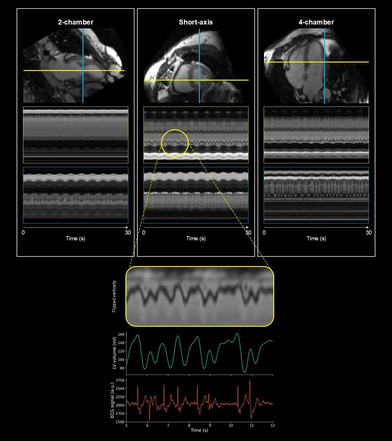
اختُبرت الطريقة على نموذج قلب اصطناعي (فانتوم)، وعشرة متطوعين أصحاء، وعشرة مرضى يعانون من انقباضات بطينية سابقة لأوانها. مع الفانتوم، تطابقت النتائج مع القياس المرجعي بشكل شبه مثالي: كسر القذف 17.86% مقابل القيمة الحقيقية 17.27%. وفي البشر، أكد تسجيل النشاط الكهربائي للقلب أن كل قفزة في الحجم تزامنت مع لحظة الاضطراب.
في السابق، كانت هذه التفاصيل مخفية في الصورة المتوسطة. الآن يمكن رؤية كيف يتعثر القلب ثم يعود إلى إيقاعه الطبيعي.
الملخص الأصلي
كما كتبه المؤلفون، بالإنجليزية arXiv:2603.04233
Conventional cardiovascular magnetic resonance (CMR) cine imaging combines data across multiple heartbeats, an assumption that can fail in arrhythmia because beat-to-beat variation causes motion artifacts and obscures functional heterogeneity. Although 2D real-time cine resolves individual beats, stacked slices are suboptimal for capturing complex 3D cardiac dynamics. We investigated continuous beat-to-beat volumetric quantification in patients with premature ventricular contractions (PVCs) using free-running 3D real-time CMR. CMR-MOTUS was extended to jointly reconstruct time-resolved 3D motion fields and a motion-corrected reference image from continuously acquired data without breath-holding or ECG gating. Data were acquired with a variable-density Cartesian trajectory and either 3D spoiled gradient-echo or balanced steady-state free-precession imaging. Ventricular volumes were obtained by propagating one manual reference-image segmentation through all reconstructed frames. The method was evaluated in a cardiac motion phantom with static ground-truth acquisitions, 10 healthy volunteers, and 10 patients with PVCs; all in vivo scans were performed after contrast administration. Phantom ejection fraction (EF) agreed closely with ground truth (17.86% versus 17.27%). Healthy volunteers showed narrow beat-to-beat EF distributions, whereas patients with PVCs showed broader and sometimes bimodal distributions. Simultaneous ECG recordings supported the temporal correspondence between volume irregularities and PVC episodes. Free-running joint 3D motion-field and image reconstruction enables continuous beat-to-beat volumetric assessment and can reveal functional heterogeneity obscured by gated or heartbeat-averaged methods. Larger studies are required to establish clinical validity and determine its role alongside standard 2D cine analysis.
كسر القذف هو نسبة الدم التي يضخها القلب في كل انقباض. عند الشخص السليم تبلغ حوالي 55–70%، بينما في الفانتوم المستخدم في هذه الدراسة بلغت 17.86% فقط.
في فيلم «تذكر كامل» (Total Recall)، يُمسح دماغ البطل في الزمن الحقيقي لرؤية ذكريات مخفية. الفكرة هنا مشابهة: بدلاً من الصور المتوسطة، تصوير متواصل لعضو حي، حيث يظهر كل خلل بوضوح.