وميض في حُلّة جديدة: الضوء أصبح أكثر سطوعًا بأربعة أضعاف سريع
وُضعت بلورة بحجم علبة كبريت تحت شعاع مسرّع سريري. غُطي نصف سطحها بنمط من نتوءات نانوية — متناهية الصغر، أصغر من طول موجة الضوء. أما النصف الآخر فتُرك أملس. كان الفرق مذهلاً: الجزء المغطى أصدر إشارة أكثر سطوعًا بمقدار 4.1 مرة.
في العلاج الإشعاعي، من المهم معرفة كمية الطاقة التي تصل إلى الورم والأنسجة السليمة بدقة. تحوّل الومّاضات الإشعاع إلى ضوء مرئي، لكن جزءًا منه يضيع داخل البلورة. يعمل البنية النانوية كعدسة صغيرة: تساعد الضوء على الخروج بدلاً من أن ينعكس مرة أخرى. اختبر الباحثون — الطلاء لم يشوّه خطية الاستجابة ولم يُدخل اعتمادًا على معدل الجرعة. بقيت البلورة مقياس جرعات أمينًا.
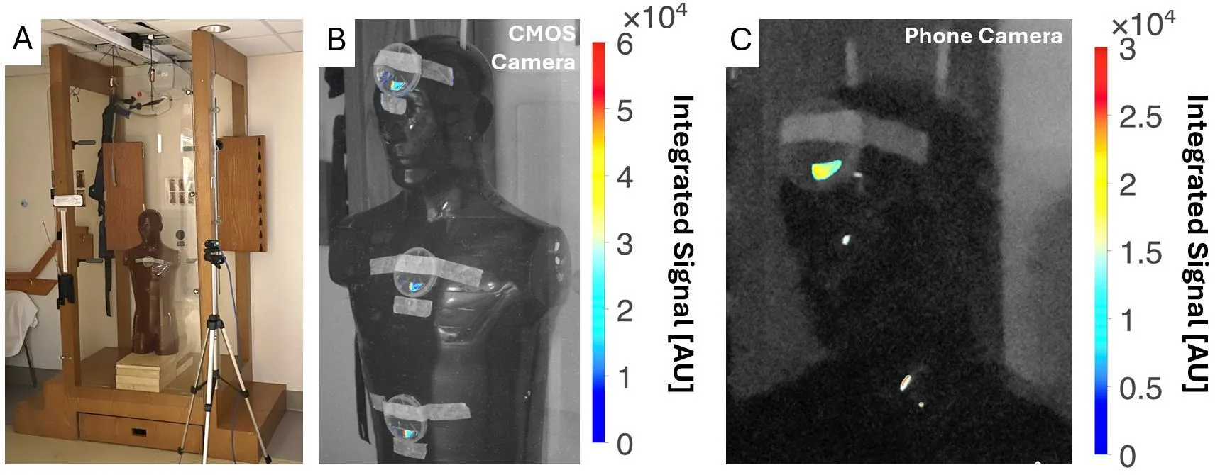
الاختبار النهائي أُجري على دمية في غرفة التشعيع الكلي للجسم. كان الضوء من النصف النانوي الفوتوني مرئيًا بوضوح على كاميرا علمية من نوع CMOS وعلى هاتف ذكي عادي — حتى مع إضاءة الغرفة. النصف الأملس ظل صامتًا. كانت الحساسية كافية لرؤية الإشارة دون أجهزة معقدة.
الملخص الأصلي
كما كتبه المؤلفون، بالإنجليزية arXiv:2606.16007
Purpose: Recent advances in metasurface photonics and manufacturing have enabled a nanophotonic surface coating to be applied to conventional scintillators, which has been shown to significantly improve light yield. However, the dosimetric properties of such coatings has not been established. We performed the first dosimetric characterization of a nanophotonic scintillator and explored clinical application to real-time in-vivo total body irradiation (TBI) dosimetry. Methods: A 4.5x1.5 cm cerium-doped yttrium aluminum garnet (YAG:Ce) scintillator, half patterned with the nanophotonic structure, half left unpatterned, enabled direct comparison between conventional and nanophotonic surfaces. The scintillator was placed in a 3D-printed light-tight box with an off-axis CMOS camera and irradiated with a clinical linear accelerator. Absolute dosimetry was done with dose-calibrated radiochromic film. For TBI, the scintillator was positioned on an anthropomorphic phantom in a TBI booth, with signal measured by both CMOS and consumer-grade phone cameras under different room lighting conditions. Results: The nanophotonic scintillator showed a 4.1x increase in signal and a 3.7x increase in contrast-to-noise ratio versus the conventional scintillator. Both scintillators exhibited dose-rate independence and linear dose response, with modest energy dependence. For TBI, the nanophotonic scintillator produced a clearly detectable signal with both CMOS and phone cameras, whereas the conventional scintillator signal was undetectable. Conclusions: Nanophotonic structures significantly enhance light output of conventional scintillators without impacting their dosimetric properties. Nanophotonic scintillators may enable real-time in-vivo TBI dosimetry. Future work should investigate this technology for improved dosimetry equipment and X-ray imaging detectors.
الطلاء القائم على الأسطح الفوقية يعمل مثل الطبقة المضادة للانعكاس على النظارات: يخمد الانعكاسات غير المرغوب فيها ويسمح بمرور المزيد من الضوء.