ماسح ضوئي يرى الورم من الداخل سريع
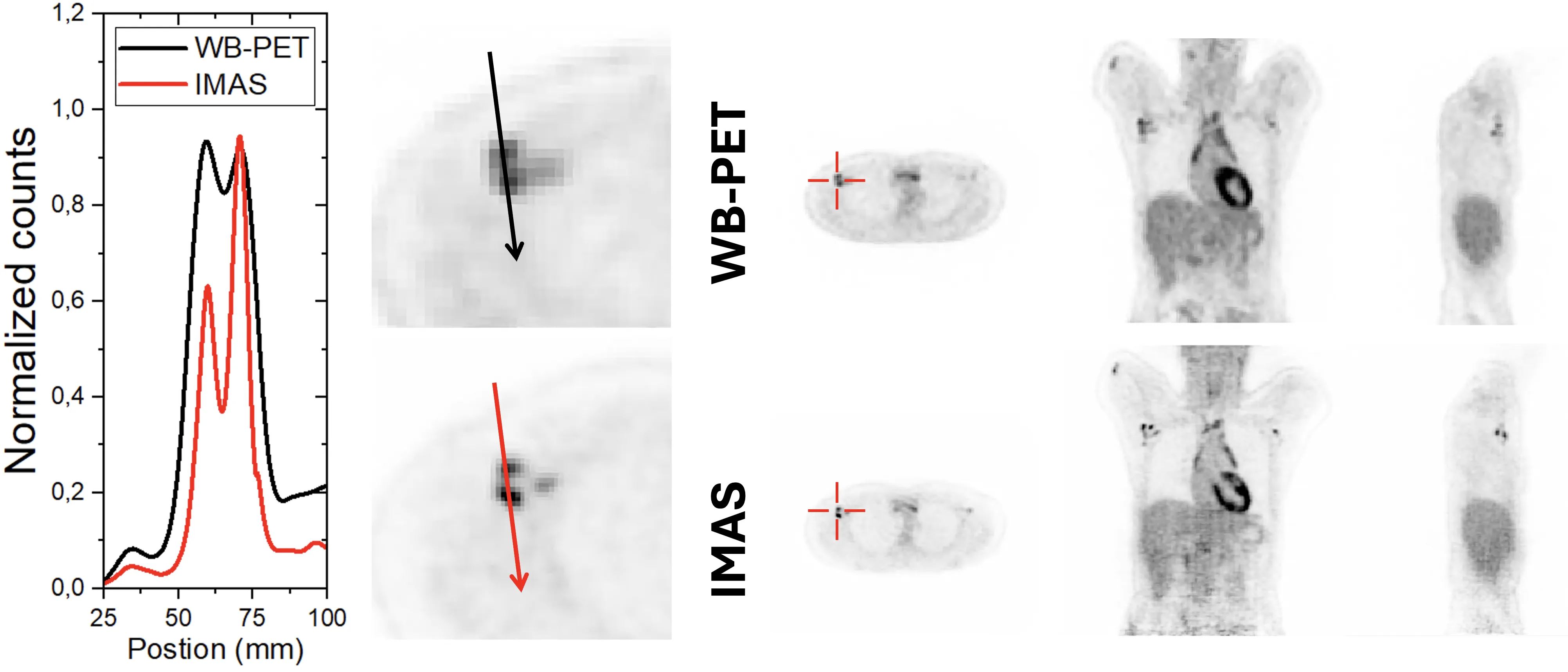
الماسح الضوئي التقليدي للتصوير المقطعي بالإصدار البوزيتروني (PET) يشبه كاميرا بعدسة ضيقة: لتصوير الجسم بالكامل، يجب تحريكه ذهابًا وإيابًا والتقاط العديد من اللقطات. أما الماسح الجديد IMAS فهو كاميرا واسعة الزاوية تغطي 71 سنتيمترًا من الجسم دفعة واحدة. يستلقي المريض بلا حراك، ويجمع الجهاز صورة من قمة الرأس إلى الفخذين في مسح واحد.
داخل الماسح توجد حلقات من بلورات شبه شفافة تلتقط ومضات الضوء الصادرة عن علامة مشعة تُحقن في الدم. كل ومضة هي إشارة من اضمحلال بوزيترون، وكلما حدد الجهاز بدقة أكبر مكان وزمان نشأتها، كانت الصورة أوضح. يجمع IMAS لأول مرة بين أسلوبين: حساب زمن طيران كمات غاما وقياس العمق الذي تمتص فيه داخل البلورة. سابقًا، كانت هاتان الإمكانيتان متاحتين بشكل منفصل فقط.
أظهرت التجارب الأولى على المرضى أن الجهاز يرى الورم بشكل أفضل من ماسح PET/CT التجاري. ومع ذلك، فإن سرعة جمع البيانات لا تزال أقل من المتوقع — بسبب عنق زجاجة في نقل المعلومات إلى الكمبيوتر. يعرف المهندسون بالفعل ما يجب إصلاحه: استبدال قناة الاتصال، وسيعمل الماسح بكامل طاقته.
الملخص الأصلي
كما كتبه المؤلفون، بالإنجليزية arXiv:2603.09898
This work summarizes the design, construction, initial performance evaluation and pilot clinical results of the IMAS system, a long axial field of view (FOV), also known as total-body (TB-), positron emission tomography (PET) prototype scanner. This PET enables for the first time in TB-PET imaging, simultaneously time-of-flight (TOF) and depth-of-interaction (DOI) capabilities. The IMAS detector block is based on LYSO semi monolithic scintillators, with individual slab sizes of 3 mm x 25 mm x 20 mm each. Arrays of 1x8 slabs are coupled to 8x8 Silicon Photomultiplier arrays. A proprietary readout reduces the 64 signals to only 16 outputs, preserving both 3D photon impact positioning and timing accuracy. IMAS has a total of 30,720 channels. PETsys electronics is used for data acquisition. The IMAS geometry is based on 5 rings of 10 cm each, with a 5 cm gap between them. It defines an axial FOV of 71 cm with a bore aperture of 82 cm. We report in this work the pilot tests of the system performance and the first clinical results. We found that the system spatial resolution remained below 4 mm across the entire FOV, even at the off-radial position of 30 cm. A coincidence time resolution with a small size 22Na source of 560 ps FWHM was measured. A sensitivity of 56.54 cps/kBq is in good agreement with previous simulation studies; however, the noise equivalent count rates performance (79 kcps at 3.26 kBq/mL) was significantly lower than expected, likely due to a data transfer bottleneck between the system and the acquisition workstation. Finally, a comparison of one of the imaged patients with a commercial TOF PET/CT scanner is also provided, pinpointing an improved tumor identification for IMAS, and the advantages of TOF and especially DOI capabilities.
كاشفات IMAS مبنية من بلورات تشبه طوبًا شبه شفاف بأبعاد 3×25×20 ملم. كل طوبة هي بمثابة عدسة مصغرة لا تلتقط الضوء فحسب، بل تخبرنا أيضًا عن العمق الذي جاء منه.