كيف ينقبض المثانة دون صبغة سريع
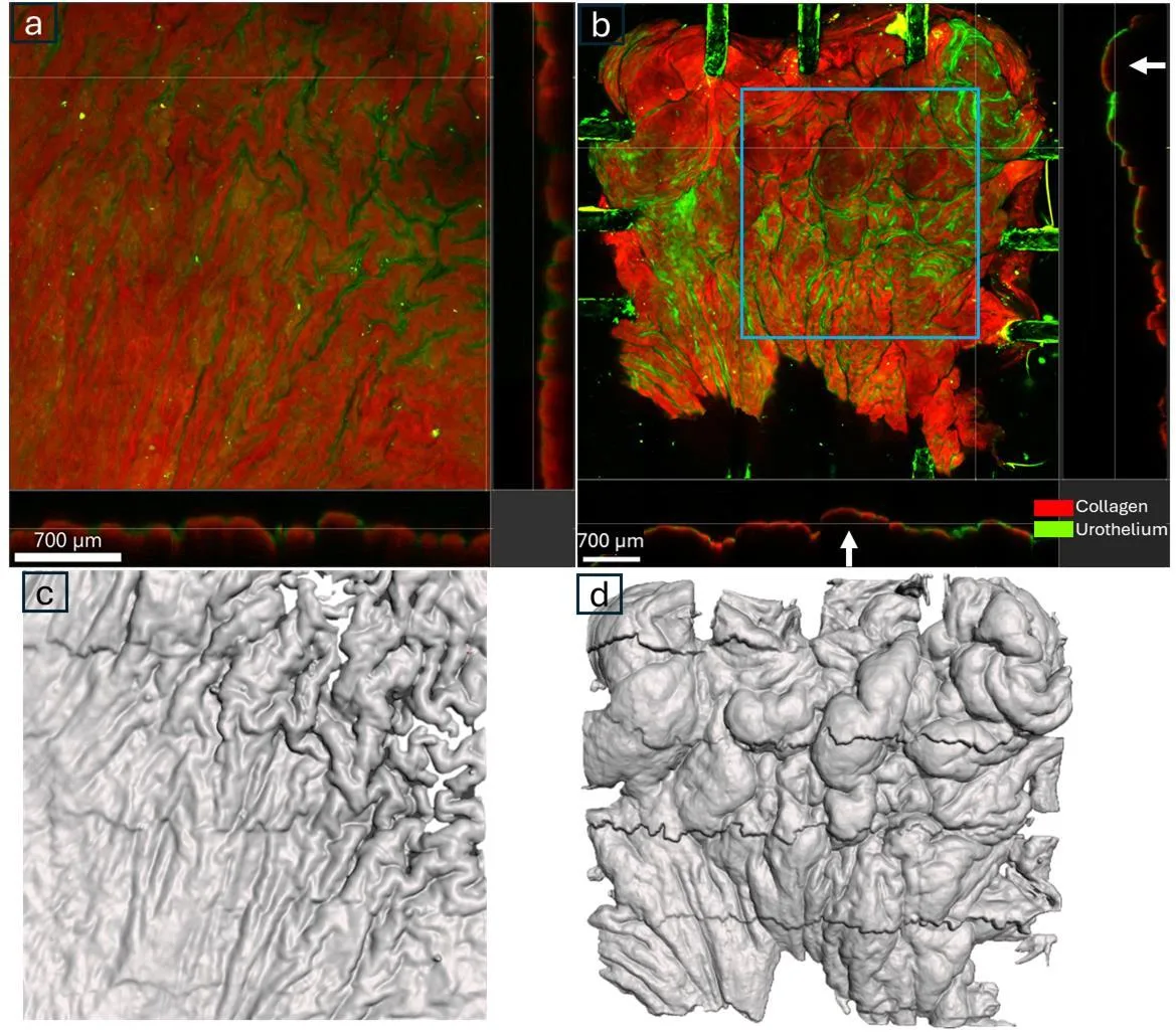
جدار المثانة ليس كالبالون المطاطي. فعندما يفرغ العضو، يتجعد سطحه مثل ورقة مكومة، ويكون التوتر مختلفًا من مكان لآخر. في السابق، لقياس ذلك، كانت توضع نقاط صغيرة ملونة على النسيج. لكن الصبغة بحد ذاتها تغيّر سلوك النسيج الحي — تمامًا كطبقة إضافية من الشريط اللاصق على بالون.
الطريقة الجديدة تستغني عن الصبغة. كاميرا تلتقط النمط الطبيعي للسطح الداخلي للمثانة، وبرنامج CoTracker3 يتتبع كل منطقة صغيرة بدقة، مثل خبير آثار يتتبع الآثار على الرمال. اختُبرت الطريقة على أربع جرذان: أثناء الانقباض، كانت المثانة تنضغط طوليًا أكثر من عرضيًا. وتحت المجهر، شوهدت طيّات كبيرة تتشكل.
هذه النظرة غير التلامسية مفيدة ليس فقط للمثانة. فأي مادة تنكمش أو تتمدد أو تغير شكلها يمكن دراستها بدقة أكبر — من عضلة القلب إلى غلاف التغليف.
الملخص الأصلي
كما كتبه المؤلفون، بالإنجليزية arXiv:2601.01315
Accurate quantification of local strain fields during bladder contraction is essential for understanding the biomechanics of bladder micturition, in both health and disease. Conventional digital image correlation (DIC) methods have been successfully applied to various biological tissues; however, this approach requires artificial speckling, which can alter both passive and active properties of the tissue. In this study, we introduce a speckle-free framework for quantifying local strain fields using a state-of-the-art, zero-shot transformer model, CoTracker3. We utilized a custom-designed, portable isotonic biaxial apparatus compatible with multiphoton microscopy (MPM) to demonstrate this approach, successfully tracking natural bladder lumen textures without artificial markers. Benchmark tests validated the method's high pixel accuracy and low strain errors. Our framework effectively captured heterogeneous deformation patterns, despite complex folding and buckling, which conventional DIC often fails to track. Application to in vitro active bladder contractions in four rat specimens (n=4) revealed statistically significant anisotropy (p<0.01), with higher contraction longitudinally compared to circumferentially. Multiphoton microscopy further illustrated and confirmed heterogeneous morphological changes, such as large fold formation during active contraction. This non-invasive approach eliminates speckle-induced artifacts, enabling more physiologically relevant measurements, and has broad applicability for material testing of other biological and engineered systems.
مثانة الجرذ بحجم حبة البازلاء، لكن جدارها عند الانقباض يشكل طيّات تشبه التضاريس الجبلية مصغرة.