خريطة عمر الدماغ سريع
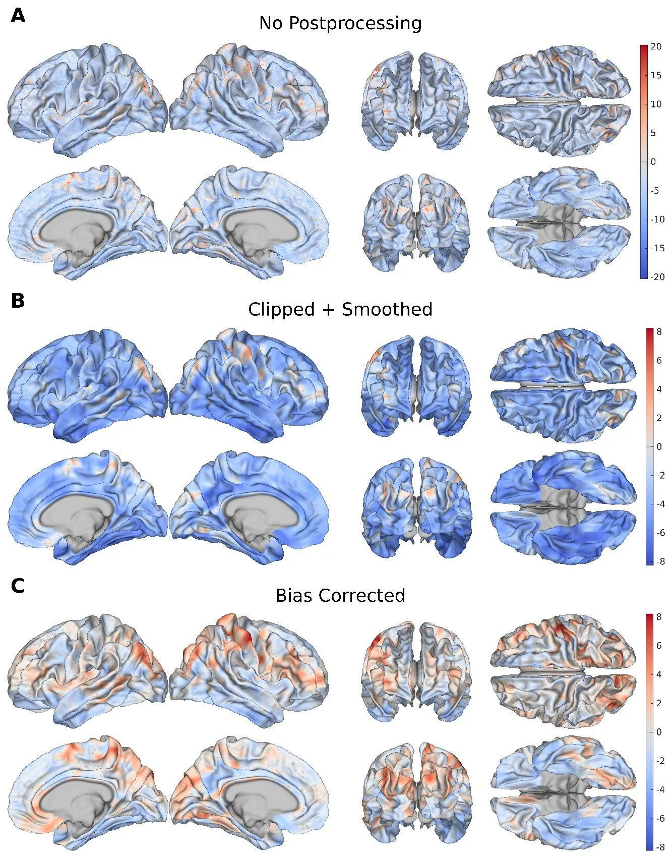
تشبه القشرة الدماغية ورقة مجعّدة: لها سُمك وانحناءات وعمق للطيات. مع التقدم في العمر، تتغير هذه الورقة بشكل غير متساوٍ — في بعض الأماكن تزداد رقة، وفي أخرى تفقد وضوح انحناءاتها. سابقًا، كان الأطباء يقيّمون عمر الدماغ ككل، وكأنهم ينظرون إلى الورقة من بعيد. أما الآن، فتدقق الشبكة العصبية في كل نقطة على السطح بخطوة مقدارها 1.37 ملم — مثل عدسة مكبرة تُظهر شيخوخة المناطق الفردية.
دُرّبت النموذج على صور لـ 14,423 شخصًا بالغًا أصحاء. ووجدت أنه لدى الأصحاء، تشيخ أسرع المناطق التي تربط أجزاء الدماغ المختلفة — المناطق الترابطية في القشرة (association areas). وفي حالات ضعف الذاكرة الخفيف، ينتشر الشيخوخة على نطاق أوسع، ليشمل التلفيف المجاور للحُصين (parahippocampal gyrus) — وهي منطقة مهمة للتذكر. وفي مرض الزهايمر، تغطي التغيرات المرتبطة بالعمر القشرة بأكملها تقريبًا، وخاصة الفصين الصدغيين.
الفرق بين العمر الحقيقي للدماغ والعمر المتوقع مرتبط باختبارات الذاكرة والتفكير. فكلما بدت منطقة من القشرة "أكبر سنًا"، كان أداء الشخص أسوأ في المهام. وهذا يعني أن خريطة الشيخوخة الموضعية ليست مجرد صورة، بل أداة للتشخيص المبكر.
تنظر الشبكة العصبية إلى القشرة كما لو كانت شبكة طرق: كل نقطة مرتبطة بجاراتها، وشيخوخة منطقة واحدة تسحب معها مناطق أخرى. هذا المنهج يتيح رؤية مكان بدء التدهور قبل ظهور الأعراض الواضحة بوقت طويل.
الملخص الأصلي
كما كتبه المؤلفون، بالإنجليزية arXiv:2601.10912
Estimating brain age (BA) from T1-weighted magnetic resonance images (MRIs) provides a powerful framework for quantifying anatomical brain aging. Whereas global BA (GBA) summarizes overall brain health, local BA (LBA) provides cortically specific patterns of aging at the subject level. Although previous studies have examined anatomical contributors to GBA, to our knowledge, no framework has been established to estimate LBA using cortical morphology. To address this gap, we introduce a graph neural network (GNN) that uses morphometric features$$\unicode{x2013}$$cortical thickness, surface area, curvature, gray/white matter intensity ratio (GWR), sulcal depth$$\unicode{x2013}$$to estimate LBA across the cortical surface at high spatial resolution (mean inter-vertex distance = 1.37 mm). Trained on cortical surface meshes extracted from the MRIs of cognitively normal (CN) adults (N = 14,423), our model achieves lower mean absolute error (MAE) than the existing state-of-the-art while identifying more biologically plausible patterns of aging in Alzheimer's disease (AD) on the ADNI dataset. Association cortices emerge as primary sites of morphometric aging in CNs, whereas mild cognitive impairment is characterized by widespread aging that is pronounced in the parahippocampal gyrus. AD subjects demonstrate significant aging across the entire cortex, particularly within medial temporal regions and associated cortical networks. Feature ablation highlights curvature and GWR as preferentially sensitive to AD pathology. Regional LBA gaps are significantly associated with neuropsychological measures of AD-related cognitive impairment, linking cortical aging patterns to clinical outcomes. These results demonstrate that GNN-based modeling of cortical morphometry enables biologically interpretable mapping of local brain aging with greater interpretability than prior work.
متوسط المسافة بين النقاط على خريطة القشرة هو 1.37 ملم، وهو أرق من عود الثقاب.
في مسلسل «الكربون المعدَّل» (Altered Carbon)، كان يمكن قراءة عمر القشرة الدماغية مباشرة — وهذا العمل يقرّبنا من مثل هذا التشخيص.