How a Tendon Tears from Within express
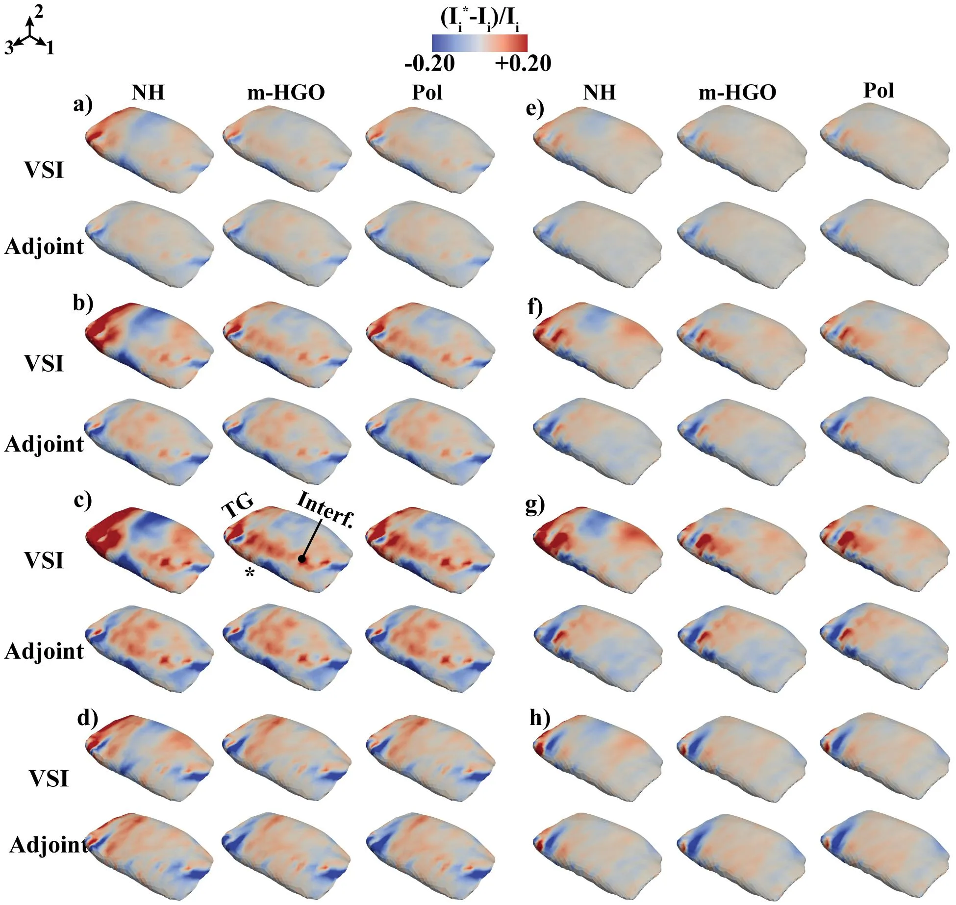
The shoulder tendon is like a cable that holds your arm and lets it rotate. When that cable gets injured, its internal structure changes, and it starts to stretch differently. In this study, they took a rat's supraspinatus tendon and pulled it along its fibers while recording how every point inside it shifted. The result was a complete 3D picture of deformation — for both healthy and injured tendons.
Next came the question: how to describe this behavior with equations? The scientists tested three models of varying complexity. The first, simplest one treated the tissue as a uniform rubber. The second, more sophisticated, accounted for fibers that resist stretching differently along and across. The third was even simpler than the second but included a term for shear. It turned out the simplest model fails: it misses how the injured tendon starts to shear internally. The middle and simplified models performed equally well, even though one uses more parameters.
Why does this matter? If we can accurately predict how a tendon will behave after injury or surgery, doctors can tailor treatments individually. For now, no model captures every nuance, but even the simplified version picks up the main trends. It's like a rough map of the terrain: you won't navigate every rock with it, but it points you in the right direction.
An injured tendon is like a rope where some strands have frayed: when pulled, the remaining fibers slide and shift more than they would in an intact rope. That shear turned out to be key — the simplest model misses it, but more realistic ones catch it.
The supraspinatus tendon is one of the most commonly injured tendons in humans: it accounts for up to half of all shoulder pain.