Сцинтиллятор в новой упаковке: свет стал ярче в четыре раза экспресс
Кристалл размером со спичечный коробок поместили под луч клинического ускорителя. Половину его поверхности покрыли узором из нановыступов — крошечных, меньше длины волны света. Вторую половину оставили гладкой. Разница оказалась разительной: покрытая часть выдала сигнал в 4,1 раза ярче.
При лучевой терапии важно точно знать, сколько энергии получила опухоль и здоровые ткани. Сцинтилляторы превращают излучение в видимый свет, но часть его теряется внутри кристалла. Наноструктура работает как маленькая линза: она помогает свету выйти наружу, а не отразиться обратно. Исследователи проверили — покрытие не исказило линейность отклика и не внесло зависимости от мощности дозы. Кристалл остался честным дозиметром.
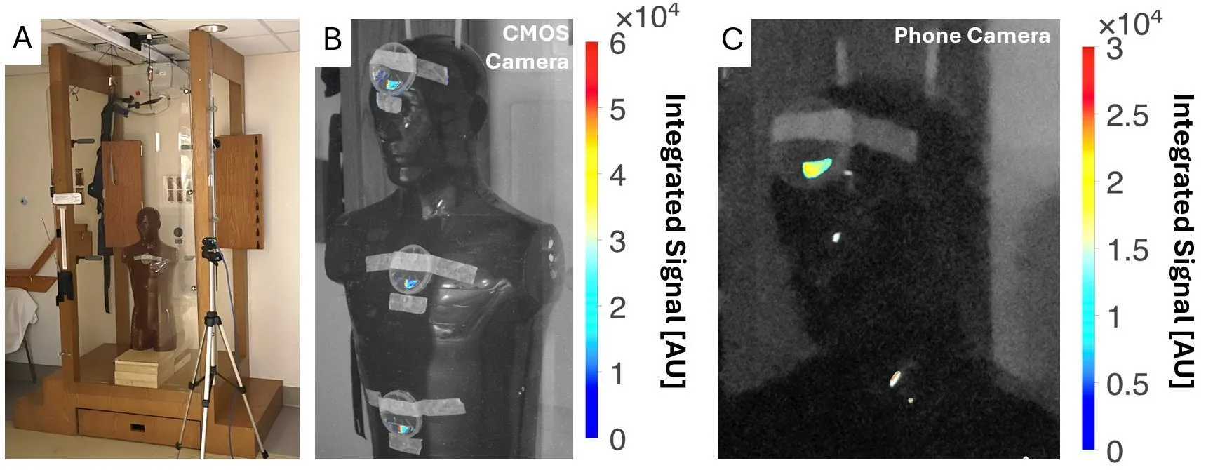
Финальный тест провели на манекене в кабине для тотального облучения тела. Свет от нанофотонной половинки был чётко виден и на научной КМОП-камере, и на обычном смартфоне — даже при включённом комнатном свете. Гладкая половина молчала. Чувствительности хватило, чтобы разглядеть сигнал без сложной аппаратуры.
Оригинальная аннотация
как её написали авторы, на английском arXiv:2606.16007
Purpose: Recent advances in metasurface photonics and manufacturing have enabled a nanophotonic surface coating to be applied to conventional scintillators, which has been shown to significantly improve light yield. However, the dosimetric properties of such coatings has not been established. We performed the first dosimetric characterization of a nanophotonic scintillator and explored clinical application to real-time in-vivo total body irradiation (TBI) dosimetry. Methods: A 4.5x1.5 cm cerium-doped yttrium aluminum garnet (YAG:Ce) scintillator, half patterned with the nanophotonic structure, half left unpatterned, enabled direct comparison between conventional and nanophotonic surfaces. The scintillator was placed in a 3D-printed light-tight box with an off-axis CMOS camera and irradiated with a clinical linear accelerator. Absolute dosimetry was done with dose-calibrated radiochromic film. For TBI, the scintillator was positioned on an anthropomorphic phantom in a TBI booth, with signal measured by both CMOS and consumer-grade phone cameras under different room lighting conditions. Results: The nanophotonic scintillator showed a 4.1x increase in signal and a 3.7x increase in contrast-to-noise ratio versus the conventional scintillator. Both scintillators exhibited dose-rate independence and linear dose response, with modest energy dependence. For TBI, the nanophotonic scintillator produced a clearly detectable signal with both CMOS and phone cameras, whereas the conventional scintillator signal was undetectable. Conclusions: Nanophotonic structures significantly enhance light output of conventional scintillators without impacting their dosimetric properties. Nanophotonic scintillators may enable real-time in-vivo TBI dosimetry. Future work should investigate this technology for improved dosimetry equipment and X-ray imaging detectors.
Покрытие на основе метаповерхностей действует подобно просветляющей плёнке на очках: оно гасит ненужные отражения и пропускает больше света.