صورة أشعة سينية بلا تشويش، كصورة فوتوغرافية بلا وهج سريع
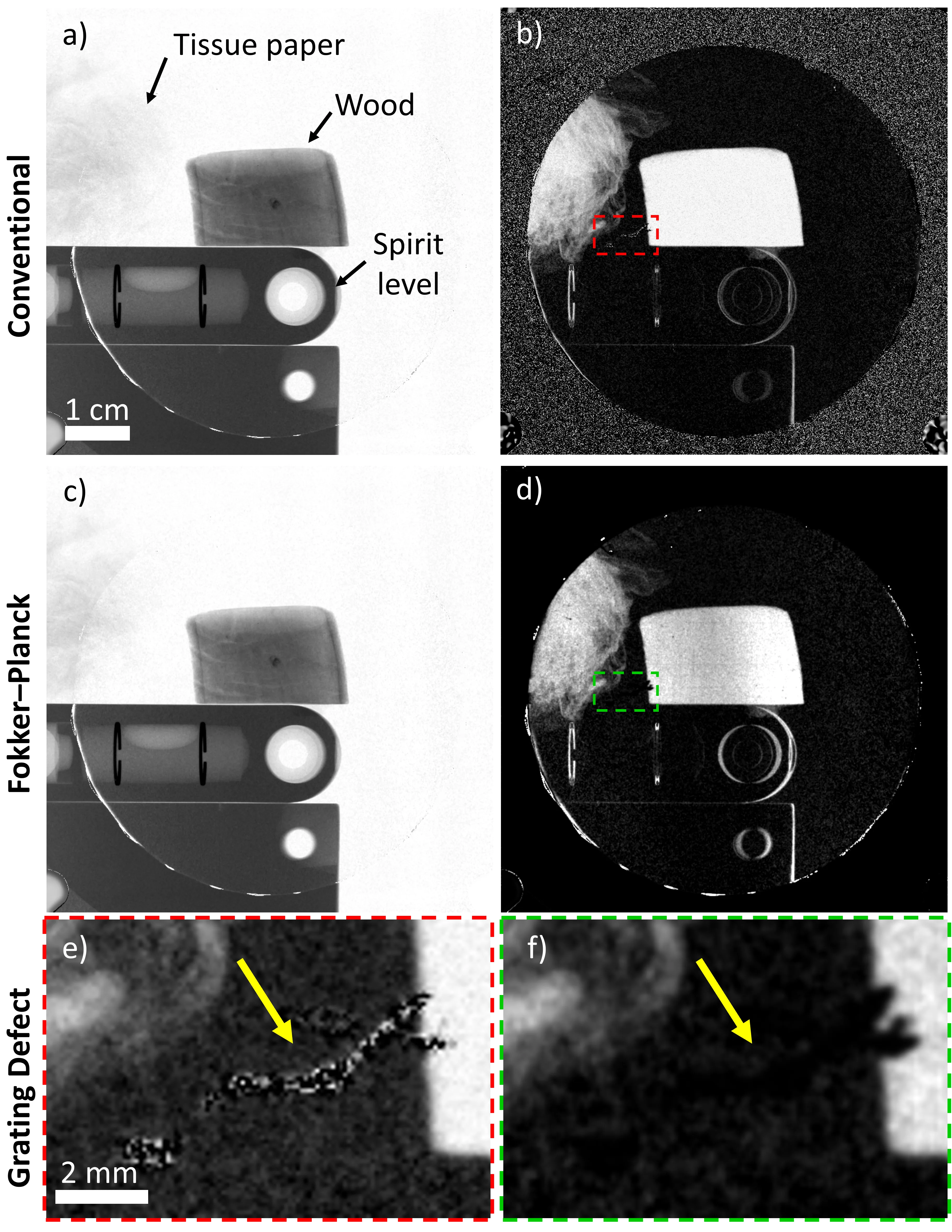
جهاز الأشعة السينية المزود بشبكات يلتقط ليس صورة واحدة بل ثلاث صور في آن واحد: صورة عادية، وخريطة لانحناءات الشعاع غير المرئية بالعين، وصورة للبنى الدقيقة التي تشتت الضوء. معًا تساعد هذه الصور في رؤية، على سبيل المثال، انتفاخ الرئة أو تليف الرئتين. لكن هناك عقبة: لفصل هذه الطبقات الثلاث، نحتاج إلى تقنية رياضية ذكية. لعقود، استُخدمت نفس الطريقة: مطابقة البيانات مع منحنى جيبي، كما لو كنا نضع موجة بإيقاع صحيح على الصورة. المشكلة أن أي خدش على الشبكة أو إشارة ضعيفة يخل بهذا الإيقاع، فتظهر على الصورة خطوط تشبه تموجات الماء عند إلقاء حجر فيه.
أخذ مؤلفو العمل الجديد معادلة تصف كيف يغير الضوء اتجاهه قليلاً ويتشتت عند مروره عبر المادة — تقريبًا مثل شعاع مصباح يدوي في الضباب. هذه معادلة فوكر-بلانك، وهي قادرة على مراعاة الصدمات العشوائية التي يتلقاها كل فوتون. بدلاً من مطابقة منحنى جيبي، تنظر الخوارزمية إلى كيفية تغير السطوع بسلاسة من بكسل إلى بكسل، وتستنتج بنفسها أين الظل الحقيقي وأين الضوضاء الناتجة عن عيوب الشبكة. النتيجة تشبه مسح الصورة بقطعة قماش ناعمة تزيل الغبار دون لمس التفاصيل المهمة.
هذا ليس مجرد تحسين تجميلي. عندما يتم التقاط الأشعة السينية بسرعة — على سبيل المثال، لتجنب طلب حبس النفس لفترة طويلة من المريض — تضعف الإشارة، وقد تخفي الضوضاء عقيدة صغيرة. الخوارزمية القائمة على فوكر-بلانك تستخرج من هذه الضوضاء معلومات أكثر فائدة من المنحنى الجيبي المعتاد. إنها مثل مرمم خبير يعيد بناء اللوحة كاملة من بضع ضربات فرشاة سليمة، دون أن يضيف من عنده شيئًا.
الملخص الأصلي
كما كتبه المؤلفون، بالإنجليزية arXiv:2605.01265
Grating interferometry is a promising diagnostic technique that enables simultaneous acquisition of three complementary, synergistic X-ray images: transmission, differential phase, and dark-field. Its key advantage over other setups is its ability to use large pixels and, hence, large-area detectors, as well as its compatibility with low-coherence, compact X-ray sources, both of which are key factors for human-scale imaging. It has already demonstrated strong potential for chest imaging applications, including the diagnosis of pulmonary emphysema, fibrosis, and cancer. To retrieve transmission, differential phase, and dark-field images from data, an algorithm is required to separate the distinct mechanisms contributing to measured contrast. Since its realization, this image-retrieval step has remained fundamentally unchanged. In this work, we develop a novel transmission- and dark-field retrieval algorithm for grating-interferometry derived from the X-ray Fokker-Planck equation. To demonstrate and validate our Fokker-Planck algorithm, we apply it to experimental measurements of a test sample and to data from a mouse chest acquired with varying exposure times and added Poisson noise. The retrieved images were qualitatively and quantitatively compared with those retrieved using a conventional sinusoidal-fitting approach. Across both samples, the Fokker--Planck method produced images consistent with conventional retrieval, with a comparable signal-to-noise ratio. Notably, our Fokker-Planck method suppresses artefacts arising in the conventional approach under grating perturbations (e.g., structural defects like scratches) and reduced flux or visibility, yielding smoother and more reproducible images. Additionally, we demonstrate that our Fokker-Planck method has an advantage over the conventional dark-field retrieval method for fast sample imaging with short exposure times and high noise.
معادلة فوكر-بلانك، التي شكلت أساس الطريقة، صُممت في الأصل لوصف الحركة البراونية — تلك القفزات العشوائية لحبوب اللقاح في الماء التي كان يراقبها تحت المجهر عالم النبات روبرت براون.
في القصص الخيالية العلمية عن ماسحات طبية مستقبلية، تتردد فكرة جهاز يرى الإنسان بوضوح تام دون أي تشويه — وهذه الخوارزمية تخطو خطوة في هذا الاتجاه.