كيف تعلّم الحاسوب قياس العضيات المتنامية سريع
ينظر عالم الأحياء إلى المجهر ليرى عشرات الكريات الخلوية الصغيرة — وهي كريات تُعرف باسم spheroids. ومن هذه الكريات ستنمو لاحقًا نماذج مصغرة للأعضاء. المهمة تشبه عمل الخباز الذي يقيّم ما إذا كانت العجينة قد ارتفعت بالتساوي في كل قالب. لكن بدل القوالب نجد الوسط الغذائي، وبدل العجين نجد خلايا حية. هذه الخلايا تتجمع ذاتيًا في كتل، وقد يكون عدد هذه الكتل في تجربة واحدة كبيرًا جدًا.
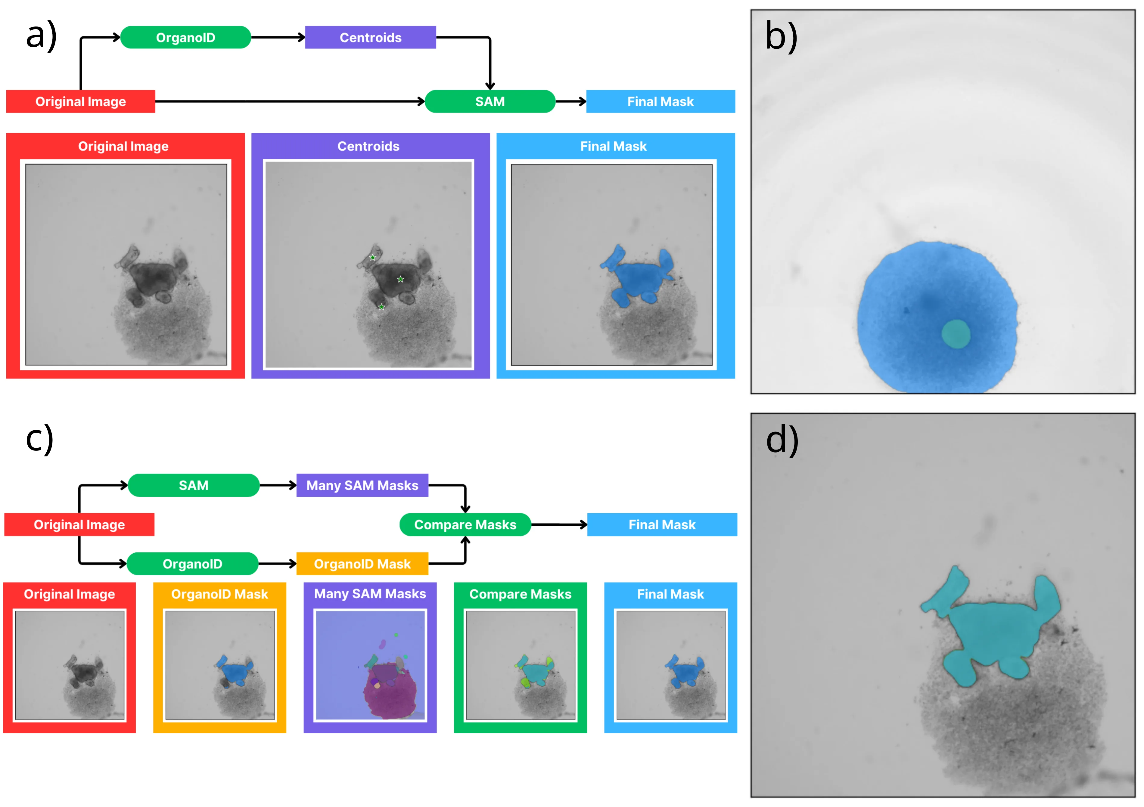
في السابق، كان العلماء يرسمون حدود كل كرة يدويًا على الصور لقياس حجمها وشكلها. كان ذلك يستغرق وقتًا طويلًا ومملًا، وكانت النتائج تعتمد على الشخص الذي يقوم بالرسم: فقد يرسم شخصان الحدود بشكل مختلف قليلًا. قرر مؤلفو الدراسة الجديدة تكليف الحاسوب بهذه المهمة الروتينية. أخذوا النموذج العام القوي Segment Anything (SAM) — وهو بمثابة «سكين سويسري» لتحديد الأجسام في أي صورة — وربطوه بأداة مصممة خصيصًا لتحليل الكريات البيولوجية. والنتيجة كانت هجينًا لا يحتاج إلى تدريب طويل من جديد.
أظهرت الاختبارات أن أياً من الأدوات الموجودة بشكل منفرد لا يعمل بشكل جيد على جميع الصور. أما الطريقة المركبة الجديدة فتعطي نتائج دقيقة ومستقرة لجميع الصور تقريبًا. حتى في أصعب الحالات — عندما تلتصق الكريات ببعضها أو تفقد وضوحها. علاوة على ذلك، فإن دقتها مماثلة لدقة شخصين مختلفين يرسمان حدود الكرة نفسها. لكن الحاسوب يفعل ذلك في ثوانٍ بدل ساعات.
الآن أصبح لدى علماء الأحياء «مقياس تلقائي» سريع وموثوق. إنه لا يتعب ولا يجادل الزملاء حول مكان رسم الخط. ستصبح مراقبة تطور الأعضاء المصغرة أسهل، والبحث عن الأدوية ودراسة الأمراض أسرع وأدق قليلًا. وهكذا، يفسح العمل الروتيني مع الحدود المجال للمراقبة الحية لكيفية بناء الخلايا لأعضاء صغيرة بنفسها.
الملخص الأصلي
كما كتبه المؤلفون، بالإنجليزية arXiv:2605.03053
Organoids are complex, three dimensional, self-organizing cell cultures which manifest organ-like features and represent a powerful platform for studying human disease and developing treatment options. Organoid development is characterized by dynamic morphological and cellular organization, which mimic some aspects of organ development. To study these rapid changes over the course of organoid development, advanced imaging and analytical tools are critical to accurately monitor the trajectory of organoid growth and investigate disease processes. In this work, we focus on computer vision and machine learning techniques to automatically measure the size and shape of developing spheroids derived from pluripotent stem cells (iPSCs), which are typically the starting material for generating organoid cultures. To facilitate this task, we introduce a composite method that combines the Segment Anything Model (SAM), a general-purpose foundation model, with an existing domain-specific tool. This composite method is evaluated together with several existing tools by testing them on organoid image data and comparing with the results of manual image segmentation. We find that no single existing tool is able to segment the test images with sufficient accuracy across all test conditions, but the newly introduced composite method produces consistent and accurate results for all but a very small fraction of the most challenging images. Finally, we compare the accuracy of this method to the variability between manual segmentations by independent annotators (inter-observer variability) and find that by one measure it performs at the level of inter-observer variability and by others it performs very close to it.
الكريات الخلوية التي تُزرع منها العضيات تشبه في شكلها وسلوكها الجنين المبكر في مراحله الأولى — ففيها أيضًا «تتفاوض» الخلايا وتنظم نفسها دون أوامر خارجية.
في فيلم «بليد رانر» تُصنع الأعضاء الاصطناعية في أنابيب اختبار — العضيات الحديثة لا يمكنها بعد استبدال كبد حقيقي، لكنها تساعد بالفعل في اختبار الأدوية دون تجارب على الحيوانات.