كيف تنظر الرنين المغناطيسي داخل الألياف العصبية سريع
الماء داخل الألياف العصبية لا يتحرك كما في كأس زجاجية، بل يصطدم باستمرار بجدران المحاور (الزوائد الطويلة للخلايا العصبية). إذا جعلنا الفاصل الزمني بين نبضات الراديو في التصوير بالرنين المغناطيسي قصيرًا جدًا، فلن يتسنى للماء الابتعاد كثيرًا، ويعكس تحركه ضيق المساحة المحيطة.
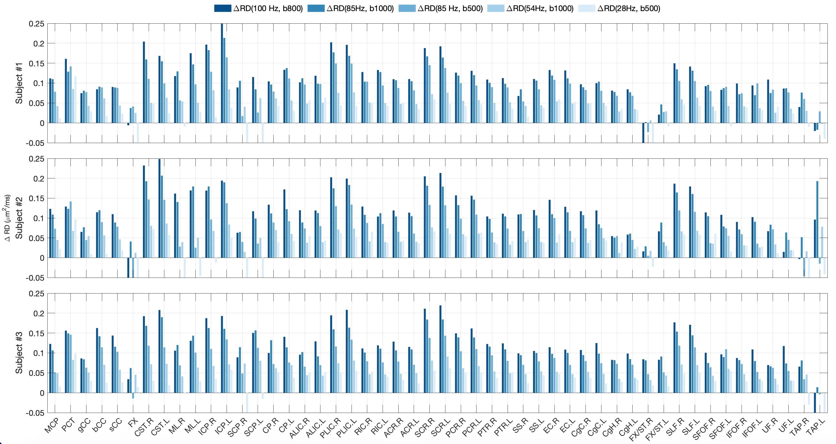
في دراسة جديدة على البشر الأحياء، قارنوا حركة الماء عبر الألياف عند زمن مراقبة قصير وآخر طويل. كان الفرق ملحوظًا بشكل خاص في السبيل القشري النخاعي، وهو الطريق الرئيسي الذي يصدر به الدماغ أوامره للعضلات. هناك تكون المحاور الأكثر سمكًا، ويستغرق الماء وقتًا أطول للانتقال من جدار إلى آخر.
سابقًا، كان يمكن ملاحظة هذا الفرق فقط على عينات الأنسجة تحت المجهر. الآن، التصوير بالرنين المغناطيسي مع التدرجات المتذبذبة يفعل الشيء نفسه دون أي شق جراحي. يعد هذا الأسلوب بأن يصبح أداة لتتبع تلف المحاور في التصلب المتعدد وغيره من الأمراض، قبل وقت طويل من ظهور الأعراض.
الملخص الأصلي
كما كتبه المؤلفون، بالإنجليزية arXiv:2607.25749
Diffusion time-dependence, defined as variations in diffusivity and/or diffusional kurtosis with diffusion time, has emerged as a valuable non-invasive imaging marker for characterizing tissue microstructural features, such as cell size, density, packing disorder, and membrane permeability. In white matter, diffusion time-dependent changes between the short diffusion time and long diffusion time in radial diffusivity (RD), defined as the diffusivity perpendicular to fiber tracts, were demonstrated to correlate strongly with mean axon diameter in ex vivo spinal cord tissues, and to reveal demyelination in mouse corpus callosum. Despite their potential to non-invasively unveil neuronal microstructures to improve the assessment and targeted therapy of neurological diseases, these novel image contrasts obtained at short diffusion times using oscillating gradient spin echo (OGSE) have only recently become feasible for human in vivo studies with high-performance gradient MRI systems. In this preliminary study, we characterized time-dependent RD with OGSE encoding in the human brain in vivo. The change in radial diffusivity between short diffusion time and long diffusion time (delta_RD) consistently exhibited high values in the corticospinal tract, indicating high sensitivity of delta_RD to large axon diameter in human brains. Imaging at a high OGSE frequency of 100 Hz and a moderate b-value of 800 s/mm2 produced the highest delta_RD in the corticospinal tract. This study established a baseline for future investigations of neuronal microstructural alterations in neurological disorders and diseases.
في السبيل القشري النخاعي، قد يصل طول المحاور إلى متر واحد، مما يجعلها أطول خلايا في جسم الإنسان.
في فيلم «ماتريكس»، كان الدماغ يُوصَّل مباشرة، لكن حتى هناك لم يكونوا يعرفون كيفية قياس سمك المحاور دون فتح الجمجمة.