مجهر أشعة سينية يرى بدقة مضاعفة سريع
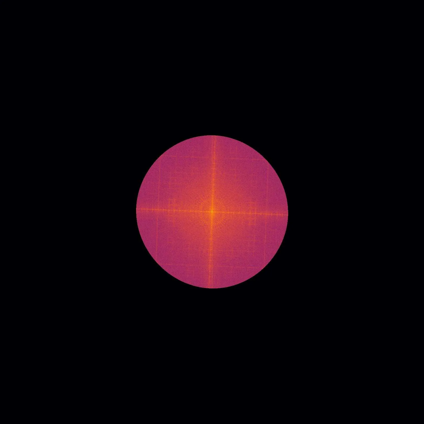
صورة الأشعة السينية للبطارية تشبه صورة فوتوغرافية من خلال زجاج ضبابي: التفاصيل الدقيقة تندمج في بقعة رمادية. السبب هو قيد الكاشف نفسه، الذي لا يميز التفاصيل الأصغر من بكسله. اقترح المهندسون تجاوز هذا الحاجز بإضاءة العينة ليس بشعاع منتظم بل بنمط مخطط.
تعمل الخطوط كاستنسل: فهي تنقل معلومات حول البنى الدقيقة إلى أجزاء الصورة التي يراها الكاشف جيدًا. وبإزاحة النمط عدة مرات والتقاط الصور، يفكك الكمبيوتر هذه البيانات ويجمع صورة بتفاصيل أصغر بمرتين من الحد السابق.
نفس المبدأ يناسب ليس فقط الأشعة السينية العادية، بل أيضًا تباين الطور والتصوير بالمجال الداكن، اللذين يظهران تفاصيل غير مرئية بالطريقة العادية. وهذا يفتح الطريق لفحص المواد والتصوير الطبي بتفاصيل كانت غير متاحة سابقًا دون أجهزة ضخمة.
الملخص الأصلي
كما كتبه المؤلفون، بالإنجليزية arXiv:2602.18343
X-ray microscopy has become an important tool for non-destructive testing, e.g., in battery research. However, imaging a cm-scale battery cell at the desired (sub-)micrometer resolution has been challenging. State-of-the-art X-ray microscopy techniques with a suited field-of-view provide (sub-) $$10\,\mu m$$ resolution, typically limited by the detector point-spread function and the (effective) detector pixel size. This work presents a super-resolution X-ray microscopy approach overcoming both limitations. It requires a structured X-ray illumination to encode high-frequency sample information that is natively unresolved within the resolved region of support. A mathematical framework is developed that decodes this information and generates a super-resolved image from multiple acquisitions with different phase shifts of the structured X-ray illumination. The presence of this encoded high-frequency information is first experimentally demonstrated, followed by quantification and validation using a resolution test chart. A resolution improvement by a factor of 2.2 is shown. Finally, we extend the proposed super-resolution technique to X-ray microtomography. Since the image acquisition scheme is inherently multimodal, phase-contrast and dark-field X-ray images can be computed additionally. These results showcase the direct impact of the proposed technique across both non-destructive testing and biomedical imaging, alleviating pixel-size limitations in detectors and sample-size restrictions.
تحسين الدقة بمقدار 2,2 مرة يعني أنه على نفس الكاشف يمكن رؤية التفاصيل التي كانت تندمج سابقًا في بكسل واحد.