A Heart Without Pauses: Movie of Every Beat express
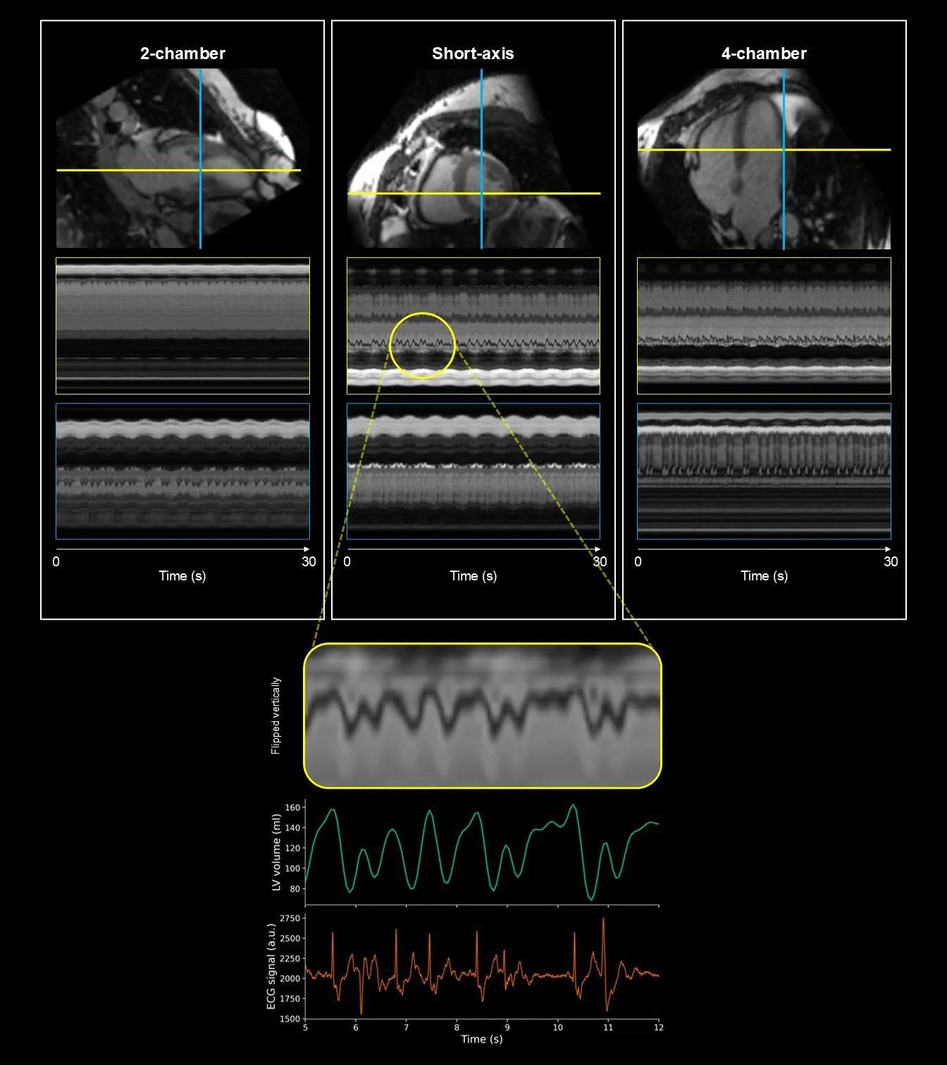
The heart beats irregularly. Standard MRI pretends all beats are the same: it stacks them on top of each other, like a photographer shooting a dancer with a long exposure. The result is a blurry smudge — pretty, but useless if you need to see each individual movement.
The new approach films the heart like a video: frame by frame, with no gaps. The patient breathes freely, and the scanner itself tracks the organ's motion and corrects the image. The doctor manually outlines the heart on one frame, and the software transfers that contour to all the others, so you can measure the ventricle's volume at every moment.
The method was tested on a heart phantom, ten healthy volunteers, and ten patients with premature ventricular contractions. For the phantom, the result matched the reference almost perfectly: ejection fraction of 17.86% versus the true 17.27%. In humans, the ECG recording confirmed that each volume spike coincided with the moment of the arrhythmia.
Previously, such details were hidden in the averaged image. Now you can see how the heart stumbles and then falls back into rhythm.
Original abstract
as written by the authors arXiv:2603.04233
Conventional cardiovascular magnetic resonance (CMR) cine imaging combines data across multiple heartbeats, an assumption that can fail in arrhythmia because beat-to-beat variation causes motion artifacts and obscures functional heterogeneity. Although 2D real-time cine resolves individual beats, stacked slices are suboptimal for capturing complex 3D cardiac dynamics. We investigated continuous beat-to-beat volumetric quantification in patients with premature ventricular contractions (PVCs) using free-running 3D real-time CMR. CMR-MOTUS was extended to jointly reconstruct time-resolved 3D motion fields and a motion-corrected reference image from continuously acquired data without breath-holding or ECG gating. Data were acquired with a variable-density Cartesian trajectory and either 3D spoiled gradient-echo or balanced steady-state free-precession imaging. Ventricular volumes were obtained by propagating one manual reference-image segmentation through all reconstructed frames. The method was evaluated in a cardiac motion phantom with static ground-truth acquisitions, 10 healthy volunteers, and 10 patients with PVCs; all in vivo scans were performed after contrast administration. Phantom ejection fraction (EF) agreed closely with ground truth (17.86% versus 17.27%). Healthy volunteers showed narrow beat-to-beat EF distributions, whereas patients with PVCs showed broader and sometimes bimodal distributions. Simultaneous ECG recordings supported the temporal correspondence between volume irregularities and PVC episodes. Free-running joint 3D motion-field and image reconstruction enables continuous beat-to-beat volumetric assessment and can reveal functional heterogeneity obscured by gated or heartbeat-averaged methods. Larger studies are required to establish clinical validity and determine its role alongside standard 2D cine analysis.
Ejection fraction is the fraction of blood the heart pumps out with each contraction. In a healthy person, it's about 55–70%, while the phantom in this study had only 17.86%.
In the movie 'Total Recall', the hero's brain is scanned in real time to reveal hidden memories. Here's a similar idea: instead of averaged snapshots, continuous imaging of a living organ, where every glitch is visible.