Computed tomography (CT) is an imaging technique that reconstructs a three-dimensional picture of an object—most commonly the human body—from a series of two-dimensional X-ray projections taken at many different angles. Unlike a single X-ray, which compresses all structures into a flat shadow, CT captures the internal geometry in slices, allowing doctors and researchers to see organs, bones, and blood vessels in their true spatial arrangement. The technique is indispensable in modern medicine for diagnosingtumors, internal bleeding, and fractures, and it has also become a standard tool in materials science and industrial inspection.
History
The mathematical foundations were laid by Johann Radon in 1917, and the first practical CT scanner was developed by Godfrey Hounsfield in the early 1970s, who shared the 1979 Nobel Prize in Physiology or Medicine with Allan Cormack.
How it works
A CT scanner rotates an X-ray source and a detector array around the object, recording hundreds of projection images. Each projection measures how much the X-ray beam is attenuated along its path. A computer algorithm—typically filtered back-projection or iterative reconstruction—solves the inverse problem: from the set of line integrals, it reconstructs the spatial distribution of attenuation coefficients inside the object. The result is a volumetric map that can be sliced in any plane or rendered as a 3D model.
In practice
CT is used routinely in hospitals for diagnostic imaging, and in industry for non-destructive testing of welds, pipelines, and electronic components.
Fun fact
The first CT scan of a human took about 4.5 minutes to acquire and required the patient to lie still in a water-filled rubber bag to reduce dynamic range.
A courtroom interpreter catches every word, every breath, every whisper in the corner. They have no right to miss a detail, because the meaning of the whole conversation depends on it. Quantum state tomography works in much the same way: it reconstructs the full quantum state of a system from a set of measurements, leaving no detail out.
A mapmaker drawing a route through a city doesn't sketch every brick in every wall. Streets, intersections, and distances are enough. The same goes for calculating radiation dose in breast tomography: you don't need every cell, just a faithful overall picture.
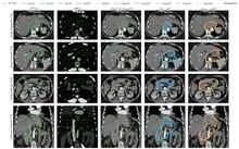
A radiation oncologist looks at an abdominal scan and mentally outlines the area to be irradiated. That area is not visible as a separate organ: its boundaries are set by protocol, not by tissue. So the doctor needs a map of the surroundings to avoid hitting healthy structures.
The nnU-Net model starts with the same task.
A globe sits on a table, and a ruler lies beside it: one is curved, the other straight. X-ray telescopes have lived with this mismatch for years. Their mirrors gather light onto a curved surface, like the globe, while the detectors that catch that light have been flat, like the ruler.
A sheet of paper left on a wet table warps and lifts its edges. No one controls that motion, but it follows the same laws as plant growth: some areas of the sheet shrink more than others, and the flat form becomes three-dimensional. Engineers have long dreamed of taming this process so that a flat blank folds itself into a desired sculpture. In the new work, that trick succeeded with accuracy down to one percent.
A deck of cards in which each card is slightly rotated cannot lie flat: the backs and faces will never align. But if the cards are flexible, they will start to adjust, shifting by fractions of a millimeter to find the perfect match. Something similar happens inside crystals when two thick layers of MoS2 or MoS2 with WS2 are stacked with a twist of 1–2 degrees.
The electron beam of a microscope usually flies through emptiness, like a courier on an empty highway. Any air molecule is a bump that makes it bounce and lose direction. So before, to examine a sample, you had to freeze it, dry it, or coat it with metal—and still you could only look in a vacuum.
A radiologist looks at an abdominal scan and sees not just organs but their spatial relationships: which kidney is larger, where the tumor lies relative to a vessel. It's like reading a map, where it's not enough to know where a city usually stands; you also need to measure the distance between two points on the ground.
In a dark room, a sunbeam passes through a pinprick in cardboard, and an upside-down image of the street appears on the wall. A pinhole camera needs no lenses: the tiny hole itself selects the rays coming from each point of the scene. Scientists at the ESRF applied this principle not to light but to X-rays, and made the camera see not shape but chemical composition.
A doctor looks at a screen and sees two images of the same part of the body. One shows where blood is rich in oxygen, the other shows how fast it rushes through the finest vessels. Previously, these images were made by different devices, and combining them was difficult. Now engineers have built a device that takes both images simultaneously.
Under an electron microscope, a protein looks like a series of blurry shadows — hundreds of thousands of images of the same molecule, but from different angles and with heavy noise. Previously, from these shadows, one first assembled a volumetric 'haze'—a map of how electric charge is distributed inside the protein.
Quantum systems made of many particles are the basis of future computers and secure networks. But the more particles there are, the harder it is to understand how they are arranged: the number of possible states grows like an avalanche. The usual approach, measuring everything, takes an incredibly long time.
A stenographer is recording a meeting, but halfway through the conversation she runs out of paper. She misses a few lines, then picks up where she left off. Later, she needs to reconstruct the full transcript without rewriting the entire conversation from scratch. That's exactly how the new method URA-SIM works for structured illumination microscopy (SIM).
A shipping container is a steel safe the size of a room, packed with goods. X-rays struggle to see through it: thick walls and dense cargo block the rays. Enter muons — particles born high in the sky that constantly rain down on Earth. They can pass through even lead, but they veer slightly off course when they encounter something heavy.
A low-dose X-ray image looks like a view through a fogged-up window: outlines are there, but details drown in a hazy veil. Doctors need such images during radiation therapy — to aim the beam precisely at the tumor and spare healthy tissue. But saving on radiation comes at the cost of noise, streaks, and artifacts that obscure what matters.
Radiation therapy is a precision strike on a tumor, but every shot must be calibrated to within a millimeter and a fraction of a second. The problem is that existing dosimeters are either bulky, or they distort the beam themselves, or they require pauses for readout.
The proton beam is like a sniper's bullet: it delivers most of its energy at a precisely set point, barely touching tissues on the way in. But over 4–6 weeks of head and neck tumor treatment, the target shifts: the patient loses weight, the tumor shrinks, posture changes. The sniper aimed at one spot, but the target has already moved.
To capture every detail of a statue's shape, you could break it into millions of tiny cubes and describe each one — slow, heavy, and your computer's memory would groan at the seams. That's how 3D medical scans are usually handled: a CT or MRI is a huge array of such cubes (voxels), and to create or improve an image, the machine has to plow through all of them.
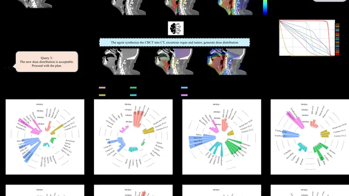
Each morning, a navigator plots a route, bypassing traffic jams and roadblocks. Similarly, before a radiation therapy session, a new AI agent recalculates the irradiation path.
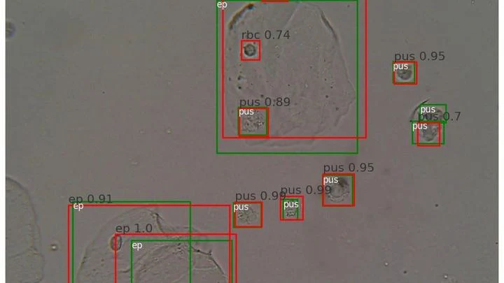
A drop of urine under a microscope isn't a flat puddle but a multi-layered volume. Like a stack of transparent pages: if you focus on one, the neighboring ones blur. The microscope's light only shows one depth layer sharply. To see all the cells, a lab technician had to take a series of snapshots, manually adjusting focus — tedious and slow.
The data from a single CT scan is comparable to several feature-length films. TCAM-Diff bypasses this bottleneck: the program compresses the 3D scene into three shadow projections — like a sculptor reconstructing a solid statue from silhouettes on three walls.
Imagine a kinked garden hose: water meets resistance, and pressure drops. A similar process occurs in arteries when they lose their straight geometry. Vascular tortuosity is not just a cosmetic defect but a factor that disrupts hemodynamics and poses a threat of ischemic stroke.
Usually a CT scanner takes many X-ray images from different angles and assembles a 3D picture. But sometimes doctors only scan a small area (say, one lung) and the data cuts off. It's like a chef given a single spoonful of soup and asked to name the exact recipe for the entire pot.
A child exploring an unfamiliar apartment crawls from object to object and remembers what is visible from each corner. This is how the brain builds a mental map — an internal model of space. The gradCSCG algorithm works on the same principle: it receives a video stream from a camera and independently draws a map of the room, relying only on the changing images.
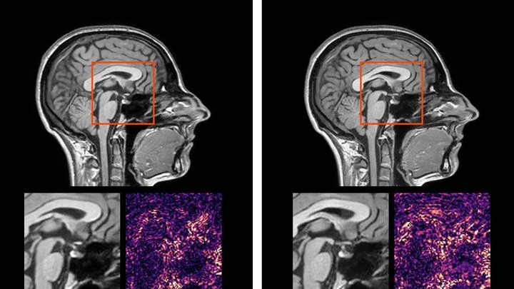
Medical scans are maps of tissue density. The brightness of each pixel reveals what’s there, much like photometry measures light intensity. But conventional algorithms analyze pixels in isolation, producing jagged organ boundaries.
A new approach compels the program to prioritize smoothness.
Imagine photographing a statue in dim light. You face a dilemma: take one bright shot or a series of dim frames from different angles. In the first case, quality is limited by photon noise, the graininess that arises from a lack of photons, while in the second, details overlap, making it harder to reconstruct the 3D structure.
Tomographs don't produce ready-made pictures. They collect numerous 'shadows' — projections of innards from different angles or using various signals — and mathematically reconstruct a 3D picture from them.
A radiologist stares at gray-white smudges on the screen — dozens of body slices where every suspicious spot must be found. Manual segmentation of one patient takes half an hour, and a large clinic sees hundreds of such patients. Fatigue and varying experience inevitably introduce variability: two specialists will outline the same lesion slightly differently.
Every second, about a hundred invisible particles, muons, pass through your body. They are born high in the sky when cosmic rays collide with air atoms, raining down on Earth. These particles are so penetrating that they can pass through a multi-story building, but they slightly change direction when they encounter dense material, such as lead or uranium.
A single camera shot stores both a normal picture and a hidden map of how light rippled through the scene. Quantum entanglement makes this possible. Pairs of linked photons are generated: one visits the object, its twin takes a detour. Later, measuring the twin one way pulls out the brightness information.
The scene is lit by spotlights, but the most interesting action happens in the shadows — where molecules invisible to the eye are hiding. The new camera picks up the heat of these "invisible" objects and turns it into ordinary light.
When you need to see inside the brain, you usually lie in a tight tube of an MRI scanner or get zapped with X-rays. Both methods are costly, bulky, and not always available.
A camera takes a snapshot, but the sensor is noisy, the optics blur, and some pixels are overexposed. Reconstructing the original picture is like guessing a melody from a scratched record: there are many possibilities, and which one is true is unclear.
A drop of blood from a vein falls under three beams, and each one drags its own clues into the light. The infrared beam makes molecules vibrate — from that trembling, you learn how much sugar and fat is in the sample. A green laser scatters off proteins and lipids, shifting color — revealing the remodeling of cell membranes.
An X-ray tomograph is like a carpenter who examines a log from all sides to see where the void is inside. It takes dozens of flat shadow images, rotating the source and detector around the object. Then a 'reconstruction' program glues these shadows into a 3D model. The quickest reconstructor is the FBP algorithm (filtered back projection).
The vessels inside the skull resemble a mountain road's hairpin turns — sharp bends, narrowings, unexpected curves. When a traffic jam forms on such a serpentine, blood stops reaching the brain properly. To predict trouble, doctors need to see the entire flow: where it accelerates, where it swirls into eddies. The full picture comes from a special MRI, like slow-motion cinematography of fluid.
A biologist looks through a microscope at dozens of tiny cell spheres — spheroids. From these, miniature organ-like structures will later grow. The task is similar to a baker's job of checking whether the dough has risen evenly in each mold. Only here, instead of molds, there's a nutrient medium, and instead of dough, there are living cells.
An X-ray machine with gratings takes not one but three pictures at once: a regular image, a map of beam deflections invisible to the eye, and an image of tiny structures that scatter light. Together, they help spot, for example, emphysema or pulmonary fibrosis. But there's a catch: to separate these three layers, you need a clever mathematical trick.
You taste a fruit smoothie through two different straws. One lets through only the big chunks, the other only the liquid. Individually, the sensations are misleading, but together they give you the key: here, it seems, is banana, and here, strawberry.
Light passing through a turbid medium like milk or living tissue scatters in all directions. Reconstructing where light was absorbed more strongly is like guessing the shape of a fallen object from a puddle on the asphalt. Optical tomography solves this problem, but usually requires large computational resources.