Acero bajo el microscopio: cómo los átomos de manganeso revelan los secretos del metal exprés
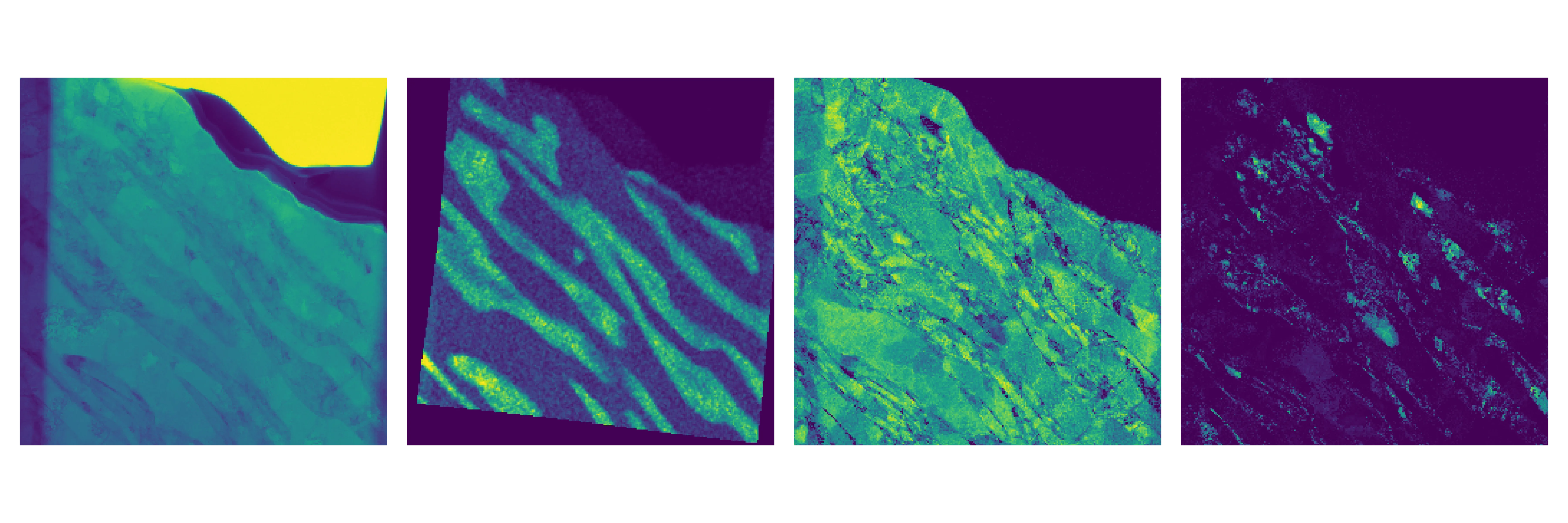
El acero no es solo un trozo de hierro. En su interior conviven diferentes estructuras, las fases, como en un pastel de capas. En el acero de ultra alta resistencia para automóviles hay tres fases: ferrita blanda, martensita resistente y austenita plástica. El acero de manganeso medio debe su fuerza precisamente a este trío. Pero cómo se distribuyen las fases en el volumen y cómo cambian bajo carga ha sido durante mucho tiempo un misterio.
Los científicos tomaron una lámina de este acero y la estiraron, como si estiraran un chicle. Luego cortaron láminas finísimas de las zonas deformada y sin deformar. Bajo el microscopio electrónico combinaron dos métodos: uno mostraba la red cristalina y el otro la distribución del manganeso. Así lograron ver por primera vez dónde está cada fase y cuánto manganeso contiene, con una precisión de 10 nanómetros, mil veces más fino que un cabello humano.
Los resultados sorprendieron: tras el estiramiento, los granos del metal se refinaron y su orientación cambió. La ferrita resultó estar menos tensionada que la martensita. La austenita se transformó parcialmente en martensita, y eso es lo que confiere al acero su ultra alta resistencia. Ahora los ingenieros podrán ajustar con mayor precisión la composición del acero para que los automóviles sean más ligeros y seguros.
Resumen original
tal como lo escribieron los autores, en inglés arXiv:2601.22407
The mechanical response of third-generation advanced high-strength steels is governed by phase transformations at the nanoscale, yet the coupled evolution of chemistry and crystallography remains poorly resolved. Here we apply a correlative scanning transmission electron microscopy approach that enables simultaneous mapping of lattice structure, crystallographic orientation, and phase distribution at 10 nanometre resolution in a medium-manganese TRIP steel. We combine nano-beam electron diffraction and energy-dispersive X-ray spectroscopy maps to characterize an industrial medium-manganese steel containing 7.15 weight percent Mn. Tensile testing of a rolled steel sample was performed, and lamellae were extracted from deformed and undeformed regions. Manganese-resolved energy-dispersive X-ray spectroscopy provides a chemical fingerprint that, when combined with nano-beam electron diffraction based phase segmentation, enables robust ferrite-martensite separation and phase-resolved lattice-parameter refinement. The phase fractions of ferrite, austenite, and martensite are quantified together with their corresponding lattice parameters, accompanied by measurable shifts in grain-size distributions and crystallographic texture in the deformed regions. Kernel average misorientation maps reveal systematically lower local misorientation in ferrite than in martensite. This multimodal workflow provides a transferable framework for quantitative, phase-resolved analysis of complex multiphase alloys at the nanoscale.
En un centímetro cúbico de este acero caben tantos límites de grano que su superficie total equivale a la de un campo de fútbol.