خريطة البلورات: كيف نرتب ملايين الصور سريع
يمسح المجهر الإلكتروني العينة ويسجل في كل نقطة كيفية تشتت الإلكترونات على الذرات. النتيجة كومة ضخمة من الصور — صورة لكل نقطة. فيها معلومات مشفرة عن ترتيب الذرات: في صفوف منتظمة أو بخلل، وإلى أين تتجه الشبكة البلورية، وهل توجد إجهادات داخلية.
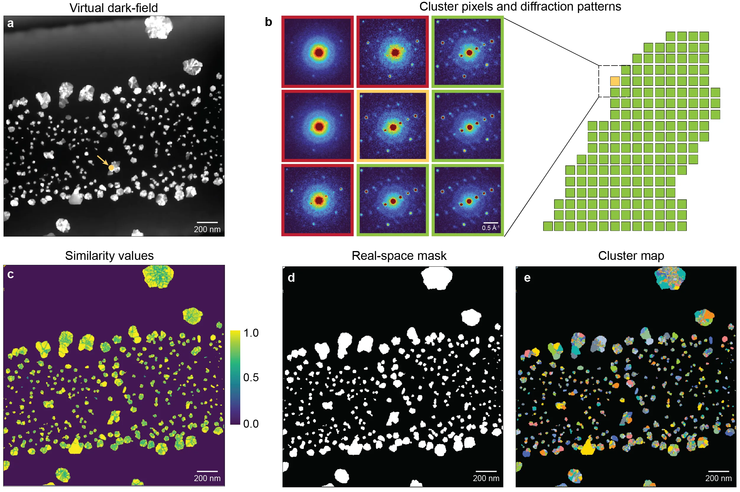
المشكلة أن هذه الصور بالملايين، ولا يمكن فرزها يدويًا. يقترح مؤلفو العمل إيجاد تلقائيًا للمناطق التي يكون فيها نمط التشتت متطابقًا تقريبًا. تُدمج الصور المتشابهة في مجموعات، ولكل مجموعة يُحصل على نمط متوسط واحد — واضح وخالٍ من الضوضاء. هكذا، من فوضى ملايين الصور تولد خريطة من عشرات المناطق.
هذا النهج يقلل حجم البيانات بشكل كبير — مئات المرات. وفي الوقت نفسه، لا تُفقد التفاصيل المهمة، بل على العكس تصبح أكثر وضوحًا: المتوسط يزيل الضوضاء العشوائية. الطريقة مفيدة ليس فقط للعينات الثابتة، بل أيضًا لمراقبة العمليات في الزمن الحقيقي، مثل نمو البلورات.
جرّب المؤلفون الخوارزمية على بيانات نمو جسيمات الذهب النانوية في وسط سائل. سجّل المجهر فيديو لتضخم الجسيمات، وقام البرنامج بتقسيم الإطارات حسب نوع الترتيب البلوري أثناء التشغيل. هذا سمح برؤية كيف تتغير بنية كل جسيم على حدة.
الآن لدى علماء المواد أداة تحول البيانات الخام من المجهر إلى خريطة واضحة: أين أي طور، وإلى أين تتجه الشبكة، وأين توجد الإجهادات. هذا يسرع التحليل ويساعد في العثور على أنماط كانت تغرق في الضوضاء سابقًا.
الملخص الأصلي
كما كتبه المؤلفون، بالإنجليزية arXiv:2601.17262
Four-dimensional scanning transmission electron microscopy (4D-STEM) enables mapping of diffraction information with nanometer-scale spatial resolution, offering detailed insight into local structure, orientation, and strain. However, as data dimensionality and sampling density increase, particularly for in situ scanning diffraction experiments (5D-STEM), robust segmentation of structurally consistent behavior across sequential measurements becomes essential for efficient and physically meaningful analysis. Here, we introduce a clustering framework that identifies crystallographically distinct domains from 4D-STEM datasets. By using local diffraction-pattern similarity as a metric, the method extracts closed contours delineating spatially contiguous regions. This approach produces cluster-averaged diffraction patterns that improve signal quality while reducing data volume by orders of magnitude, enabling rapid and accurate orientation, phase, and strain mapping. We demonstrate the applicability of this approach to in situ liquid-cell 4D-STEM data of gold nanoparticle growth. Our method provides a scalable and generalizable route for spatially coherent segmentation, data compression, and quantitative structure-strain mapping across diverse 4D-STEM modalities. The full analysis code and example workflows are publicly available to support reproducibility and reuse.
متوسط أنماط الحيود يحسن جودة الإشارة لدرجة أن انعكاسات ضعيفة تصبح مرئية، كانت غير ملحوظة في الصور الفردية.