كيف يكشف الضوء ألياف الأنسجة سريع
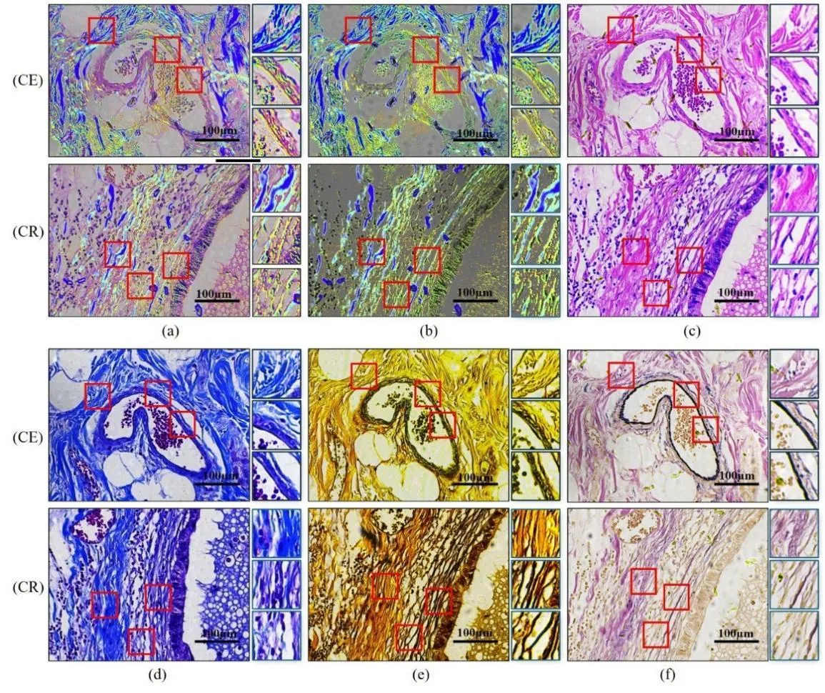
داخل أجسامنا، بين الخلايا، توجد شبكة داعمة تُعرف بالمطرس خارج الخلوي. إنها أشبه بسقالات من البروتينات والكربوهيدرات تُبقي الخلايا في مكانها. وعندما تتغير الألياف في هذه الشبكة، قد يكون ذلك علامة على مرض — مثل السرطان أو التليف.
عادةً، لرؤية هذه الألياف، يُقطع النسيج إلى شرائح رقيقة وتُصبغ بأصباغ خاصة. وهذا يستغرق وقتًا طويلًا وقد يُتلف العينة أحيانًا. في الدراسة الجديدة، جرب العلماء طريقة أخرى: فقد مرروا عبر النسيج ضوءًا مستقطبًا — وهو ضوء تهتز موجاته في مستوى واحد. الألياف المختلفة تُدير مستوى اهتزاز الضوء بزوايا مختلفة. ومن خلال تحليل هذه التغيرات، يمكن معرفة نوع الألياف التي أمامنا.
وللتمييز بين أنواع الألياف، استخدم العلماء طريقة UMAP — فهي تساعد على رؤية البنية في البيانات المعقدة، كما لو كنا نرتب الأشياء المتشابهة في أكوام مختلفة. وتبين أن الألياف الكولاجينية والمرنة والشبكية تعطي أنماط استقطاب مختلفة. وهذا يعني أنه يمكن تحديد نوع الألياف من خلال الضوء دون صبغ.
هذا النهج قد يسرّع التشخيص: بدلاً من الصبغ الذي يستغرق ساعات، نحصل على تحليل سريع تحت المجهر المستقطب. وفي المستقبل، سيكمل هذا الأساليب التقليدية ويساعد الأطباء على تقييم حالة الأنسجة بدقة أكبر.
الملخص الأصلي
كما كتبه المؤلفون، بالإنجليزية arXiv:2602.14459
Extracellular matrix (ECM) constitutes a key basement structure to human organisms by acting as a complex network of large proteins and carbohydrates that provide structural support to surrounding cells. Remodeling in the extracellular matrix's structural fibers leads to insight into the development of diseases such as cancer, fibrosis and carcinoma. While standard tissues visualization in the ECM involves multiple lengthy histopathological staining protocols, Mueller matrix-based polarimetry provides label-free tissue slices' microstructural information and optical properties. This work aims to identify three types of fiber tissues commonly found in the ECM of gastrointestinal tissue specimens by analyzing their polarization properties. To address decomposition methods' reliance on restrictive hypotheses and inability with an individual polarization-based parameter to determine the nature of a given biological tissue; this study employs Uniform Manifold Approximation and Projection (UMAP) method to offer greater discriminative power and flexibility. Subsequently, polarization-based features will be extracted and compared between fiber regions statistically to discern potential diagnostic differences. By providing colorized images, this work aims to demonstrate the feasibility of distinguishing different fibers with polarization approach, offering insights for future clinical development while complementing existing staining methods for pathological tissue specimens.
لا يُستخدم الضوء المستقطب في الفحص المجهري فقط: فالنحل، على سبيل المثال، يهتدي باستقطاب السماء حتى في الأيام الغائمة.