Рентгеновский микроскоп увидел вдвое чётче экспресс
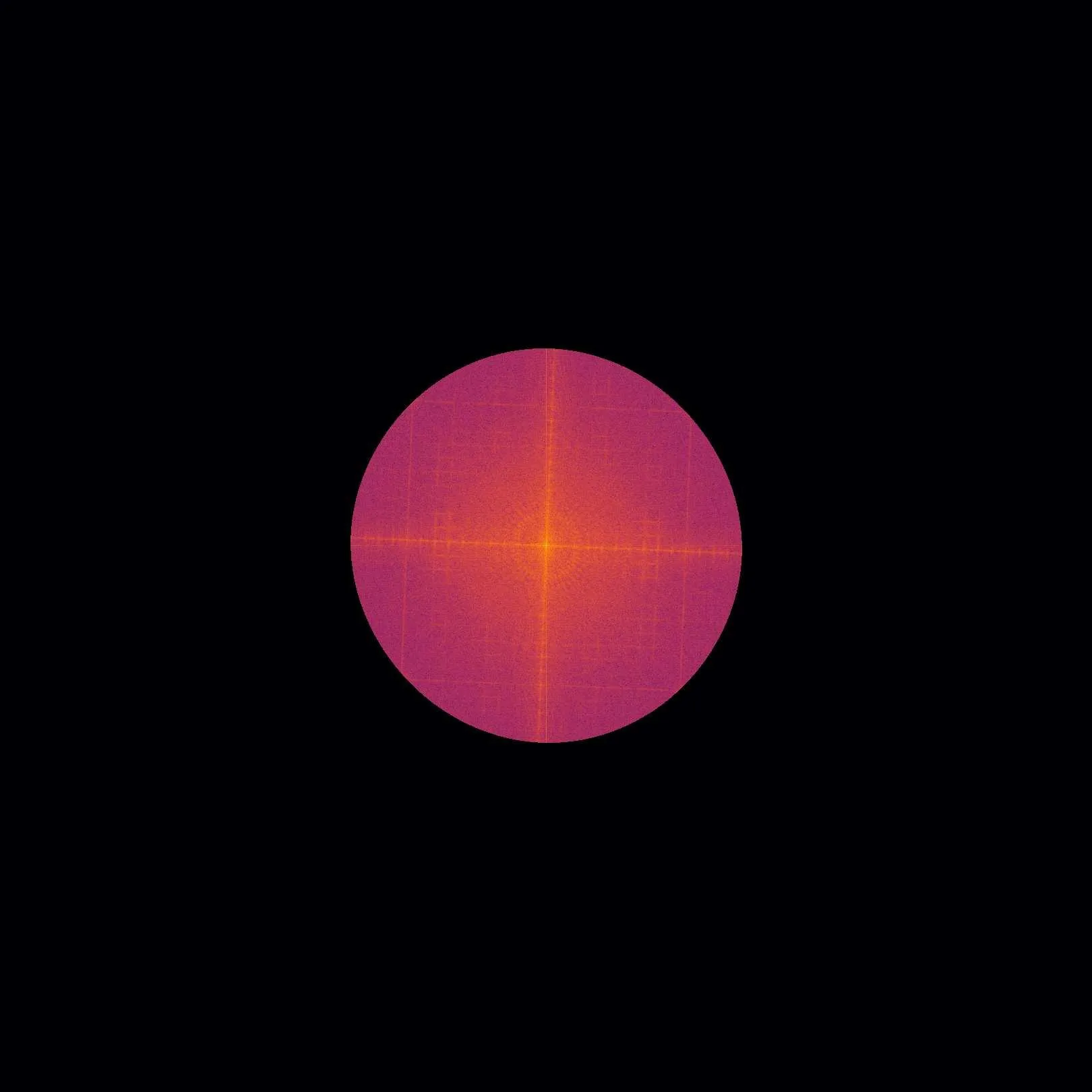
Рентгеновский снимок батареи похож на фотографию через мутное стекло: мелкие детали сливаются в серое пятно. Причина — ограничение самого детектора, который не различает детали меньше своего пикселя. Инженеры предложили обойти этот барьер, освещая образец не ровным лучом, а полосатым узором.
Полосы работают как трафарет: они переносят информацию о мельчайших структурах на те части изображения, которые детектор видит хорошо. Сдвигая узор несколько раз и делая снимки, компьютер разбирает эти данные и собирает картинку с деталями вдвое мельче прежнего предела.
Тот же принцип подходит не только для обычного рентгена, но и для фазового контраста и темнопольного изображения, которые показывают невидимые обычным способом детали. Это открывает дорогу к проверке материалов и медицинской визуализации с деталями, которые раньше были недоступны без громоздких установок.
Оригинальная аннотация
как её написали авторы, на английском arXiv:2602.18343
X-ray microscopy has become an important tool for non-destructive testing, e.g., in battery research. However, imaging a cm-scale battery cell at the desired (sub-)micrometer resolution has been challenging. State-of-the-art X-ray microscopy techniques with a suited field-of-view provide (sub-) $$10\,\mu m$$ resolution, typically limited by the detector point-spread function and the (effective) detector pixel size. This work presents a super-resolution X-ray microscopy approach overcoming both limitations. It requires a structured X-ray illumination to encode high-frequency sample information that is natively unresolved within the resolved region of support. A mathematical framework is developed that decodes this information and generates a super-resolved image from multiple acquisitions with different phase shifts of the structured X-ray illumination. The presence of this encoded high-frequency information is first experimentally demonstrated, followed by quantification and validation using a resolution test chart. A resolution improvement by a factor of 2.2 is shown. Finally, we extend the proposed super-resolution technique to X-ray microtomography. Since the image acquisition scheme is inherently multimodal, phase-contrast and dark-field X-ray images can be computed additionally. These results showcase the direct impact of the proposed technique across both non-destructive testing and biomedical imaging, alleviating pixel-size limitations in detectors and sample-size restrictions.
Улучшение разрешения в 2,2 раза означает, что на том же детекторе можно увидеть детали, которые раньше сливались в один пиксель.