Fibers That Soak Up Blood express
A drop of blood falls onto a bandage made of ultra-thin polymer fibers. Usually, it just sits on the surface like a dewdrop on wax paper. The material repels water, so the blood doesn't soak in — meaning the bandage doesn't adhere to the wound.
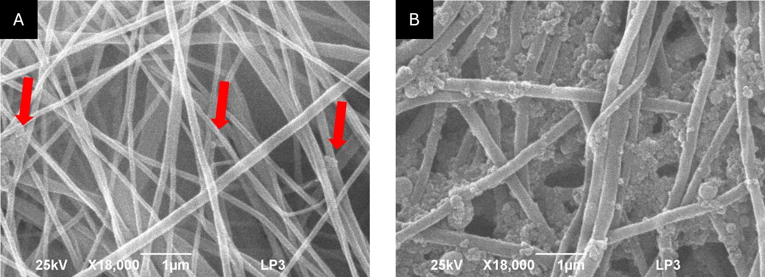
Scientists modified the fiber surface. They added tiny silicon particles to the polymer — a thousand times thinner than a human hair. The particles were made with a laser in liquid, without any adhesive additives, so they remained "bare" and active. In one version, the particles were hidden inside the fibers; in another, they were exposed on the surface, like sesame seeds on a bun.
The results were different. Fibers with particles inside only wet slightly better: the blood drop spread a bit wider but stayed compact. But when the particles stuck out, the drop flattened into a thin pancake. The contact angle dropped below 20 degrees — almost like on wet glass. Under a microscope, you could see a clear zone of plasma spreading around the red core of blood cells. The fibers, like a sponge, sucked in the liquid part of the blood, leaving the cells on top.
Such a material could become the basis for smart bandages: they quickly soak up blood, adhere tightly to the wound, and even separate blood into components. It's a step toward dressings that don't just cover a wound but actively interact with it.
Original abstract
as written by the authors arXiv:2601.05809
Electrospun poly(\epsilon-caprolactone) (PCL)nanofibrous mats are widely considered for blood-contacting wound dressings and small-diameter vascular applications; however, their intrinsic hydrophobicity limits rapid wetting and controlled interaction with blood. In this work, we modulate the interfacial response of PCL nanofibers by incorporating oxide-shelled silicon nanoparticles (SiNPs) synthesized by pulsed laser ablation in liquid, a ligand-free approach that avoids organic stabilizers and preserves surface reactivity. Two composite architectures were designed: SiNPs embedded within the fiber bulk (PAC1,4,16) and SiNPs preferentially exposed at the fiber surface (SPAC1,4,16), with systematically increasing nanoparticle loadings. Structural characterization confirmed the retention of a homogeneous fibrous morphology and the targeted nanoparticle distribution. The dynamic interaction with whole blood was quantified using time-resolved contact-angle measurements, complemented by top-view optical microscopy and three-dimensional profilometry of dried droplets. Pristine PCL remained strongly hydrophobic whereas a hydrophilic PCL functionalized with APTES showed rapid spreading. Incorporation of SiNPs within the fiber volume led to only a moderate enhancement of wettability, and dried droplets retained compact morphologies with limited spreading. In contrast, surface-decorated mats displayed a sharp, concentration-dependent transition toward highly wettable behavior: for SPAC16, the contact angle fell below 20, droplet profiles became markedly flattened, and microscopy revealed extended plasma-rich regions surrounding a red-cell-rich core, indicative of pronounced phase separation within the nanofibrous network.
A contact angle below 20 degrees means the blood drop spreads almost like water on clean glass: it becomes flat rather than dome-shaped.