How Organ Architecture Shapes Radiation Protection express
Radiation therapy targets tumors, but healthy tissue suffers too. Flash therapy delivers radiation in a fraction of a second and, remarkably, spares healthy cells without reducing the punch to the cancer. Exactly how this works remains unclear, but the authors set out to test whether this protection depends on the organ's own architecture.
Organs can be roughly divided into two types. Some are like a string of lights wired in series: one bulb burns out, and the whole string goes dark. These include the spinal cord and brainstem, and are called serial organs. Others resemble a sponge: you can cut out a piece, and the remaining part keeps working. These include the liver and lungs, and are called parallel organs. The difference matters: for a string of lights, any strong hit to a single point is dangerous, while for a sponge, only average damage across the whole volume counts.
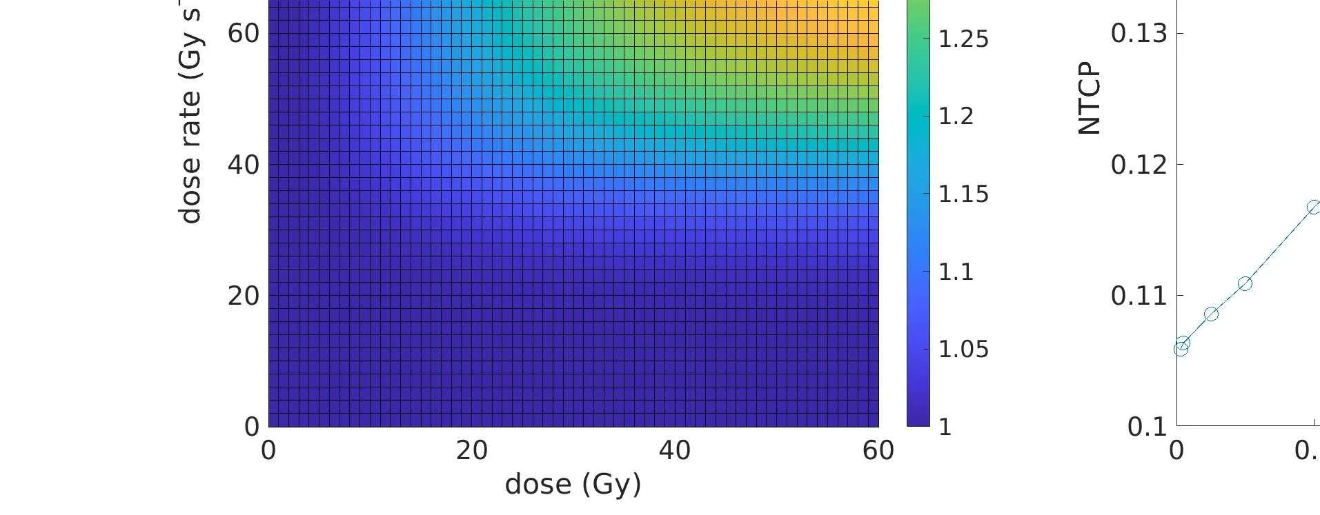
The authors built a computer model. They considered two explanations for the flash effect. The first links protection to rapid oxygen depletion in cells: radiation seems to drink up all the oxygen, making cells temporarily less sensitive. The second simply involves dose and dose-rate dependence. Both explanations assume that protection acts locally, at the site of irradiation. They then calculated the probability of complications for different organ structures under non-uniform irradiation. It turned out that for string-like organs, flash mode provides notably better protection than for sponge-like organs. For example, at the same 20% complication probability for conventional irradiation, flash reduced it to 14% for the sponge and to 11% for the string.
The conclusion is simple: if the flash effect is indeed local, then organ architecture is not just an anatomical detail but a key to predicting side effects. This means that when planning flash therapy, doctors should consider which organ lies next to the tumor: a chain or a sponge. That way, they can better protect what matters most.
Original abstract
as written by the authors arXiv:2604.25364
Objective: This study aims to investigate the influence of organ architecture (specifically the distinction between serial and parallel tissue) on the protective FLASH effect when organs are irradiated with inhomogeneous dose distributions. Approach: An in silico modeling framework was developed using two distinct methods to calculate the effective FLASH dose: the first method utilized a biophysical model of radiolytic oxygen depletion (ROD); the second employed a phenomenological logistic function where the effective FLASH dose is a function of local dose and dose rate. Both models assume that the underlying mechanism behind the FLASH effect is local. Normal Tissue Complication Probability (NTCP) for heterogeneous dose distributions was calculated using the Lyman-Kutcher-Burman (LKB) model and the generalized equivalent uniform dose, varying the volume effect parameter n from 1.0 (parallel) to below 0.01 (serial) to explore different architectures. Results: Both the ROD and phenomenological models showed FLASH sparing compared to conventional radiotherapy. Also, the sparing increased with decreasing $$n$$ (the sparing is more important for serial organs). For example, for a specific calculation, when the NTCP for conventional radiotherapy was 0.2 (set value) the corresponding NTCP for FLASH delivery ranged from 0.14 for n=1 to 0.11 for n=0.1. Significance: Our results indicate that if the underlying mechanism/s behind the FLASH effect is/are local, the toxicity sparing associated to FLASH-RT can be dependent on the architecture of the irradiated organ/tissue, being more important for serial organs, which are more sensitive to large local doses than to average doses.
Flash therapy uses doses that would be lethal if delivered over a longer time, but the ultra-short pulse makes them tolerable: it's like pouring a bucket of water over your hand in a second versus dripping it drop by drop for an hour: in the first case, the skin just gets wet, but in the second, it might become irritated.