A radioactive tracer is a substance containing a radioactive isotope that is introduced into a biological system, typically the human body, to track physiological processes or highlight specific organs for imaging. In nuclear medicine, tracers like technetium-99m or fluorine-18 are attached to molecules that accumulate in particular tissues, allowing doctors to see metabolic activity, blood flow, or tumor growth. The emitted gamma rays or positrons are detected by cameras to produce functional images.
History
The concept dates back to the 1920s with George de Hevesy, who used radioactive lead to study plant metabolism and later developed the tracer principle. Technetium-99m was introduced in the 1960s and became the most widely used medical isotope.
How it works
The tracer is injected, inhaled, or swallowed, and it travels through the body, concentrating in target organs based on its chemical properties. For gamma emitters, a gamma cameradetects the radiation and builds a 2D or 3D map of its distribution. For positron emitters (like fluorine-18), the positron annihilates with an electron, producing two gamma rays that are detected in coincidence by a PET scanner, giving precise localization.
In practice
It is used in PET and SPECT imaging to diagnose cancers, heart disease, and neurological disorders, and to monitor treatment response, such as in chemotherapy for glioblastoma.
Fun fact
The most common tracer, technetium-99m, has a half-life of about 6 hours — long enough to perform a scan but short enough to minimize radiation exposure to the patient.
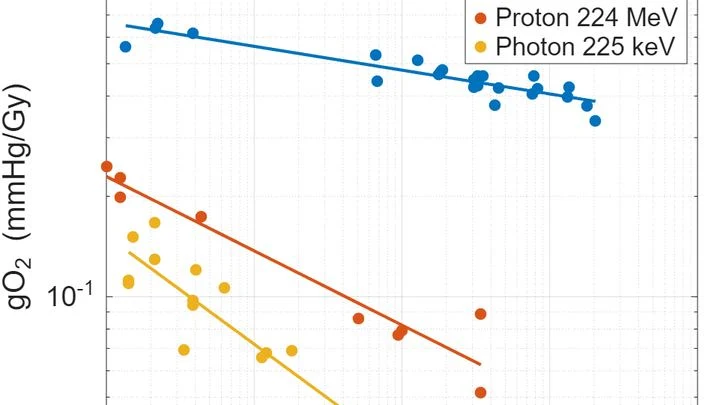
A camera flash that fires in a thousandth of a second doesn't heat the subject — unlike a regular flash that lasts a full second. A similar trick has been found in radiotherapy: when a radiation dose is delivered in milliseconds instead of minutes, healthy tissues suffer less. This is called the FLASH effect.
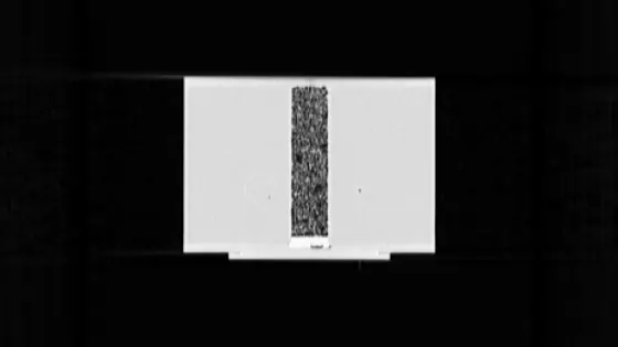
A crystal the size of a matchbox was placed under the beam of a clinical accelerator. Half of its surface was covered with a pattern of nanoscale bumps — tiny, smaller than the wavelength of light. The other half was left smooth. The difference was striking: the coated part produced a signal 4.1 times brighter.
Radiation therapy targets tumors, but healthy tissue suffers too. Flash therapy delivers radiation in a fraction of a second and, remarkably, spares healthy cells without reducing the punch to the cancer. Exactly how this works remains unclear, but the authors set out to test whether this protection depends on the organ's own architecture.
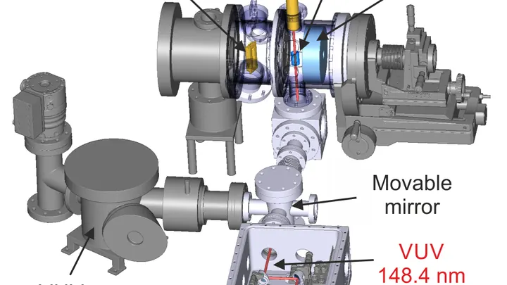
A laser beam as faint as a whisper of light has stirred a thorium-229 nucleus into a higher energy state. Most nuclei demand a blast of energy, but thorium-229 has such a tiny energy gap that even this gentle nudge works.
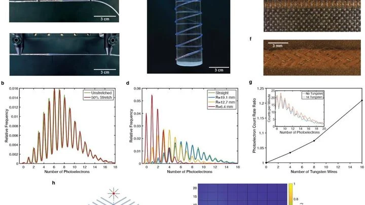
Inside an ultra-thin polymer thread are microscopic crystals — scintillators. When an invisible radioactive particle hits, they flash for a moment, like fireflies. A sensitive sensor catches each flash, turning the thread into a flexible detector.
The radioactive isotope terbium-149 acts like a tiny homing bomb: it latches onto cancer cells and detonates with alpha particles that destroy the tumor from within, barely touching healthy tissue. But this bomb has a quirk: it also glows with positrons, and that glow can be captured by a special camera.
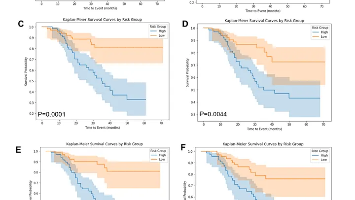
PET/CT is like a map where cancer cells light up as bright spots. But what if the map is blank and the disease is still hiding somewhere? In 132 men with recurrent prostate cancer, doctors found no glowing lesions, but they decided to study the texture of the scan itself, like an experienced tracker noticing bent grass where an animal has passed.
A beam of carbon ions strikes the tumor precisely, like a surgeon who can only see the skin. Inside the body, the beam stops, and exactly where used to be a mystery. Now, helium is mixed into the same beam: it passes through the body like an X-ray, and its trail shows where the carbon went.
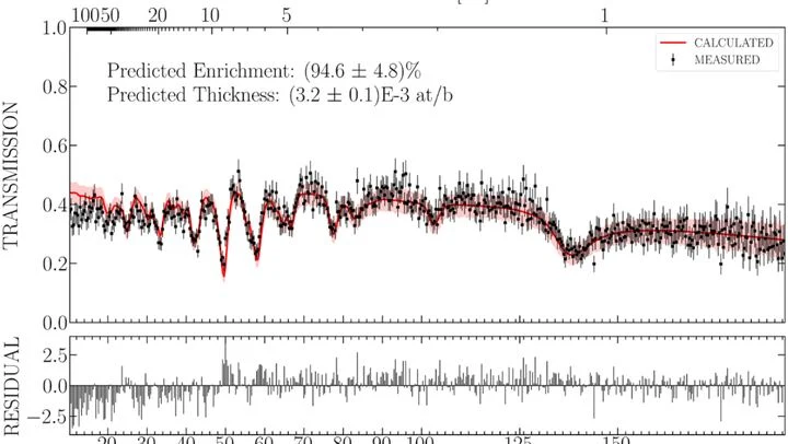
A customs officer can't take a suitcase apart to see what's inside. They shine X-rays through it and, from the shadows on the screen, tell: here's metal, here's organic matter. In much the same way, an inspector for future nuclear disarmament treaties should be able to check a warhead — without opening it or seeing its secret internals.
Neutrons are better suited for this than X-rays.
When treating epilepsy, doctors adjust the drug dose almost blindly. Too little, and seizures continue; too much, and side effects appear. Usually, a patient's blood is sent to a lab, and the result comes back a day or two later. Now, the same test can be done on a tiny silicon chip.
The sensor works like a highly sensitive nose.
Radioactive tracers are like a courier with a beacon: you inject it into the body, and it highlights the target organ on the scan. But this courier comes with strict rules: you can't send it often, it quickly 'gets tired,' and it costs a lot. Doctors have long dreamed of a way to see an organ without radiation.
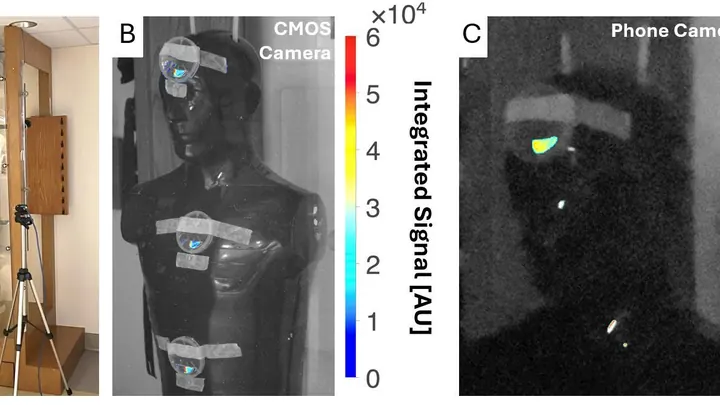
A firefighter with a thermal imager can see the source of a fire through smoke. But how do you see a radiation source that has no smell, no glow, and no heat? Gamma cameras solve this problem: they capture gamma rays emitted by radioactive materials and overlay their 'trace' onto a regular photograph. The result is an image showing that the source is, say, behind the far door.
Every year, in the United States alone, doctors perform more than 16 million scans using the radioactive isotope technetium-99m. It lights up the heart, bones, and lungs in images, helping to find tumors or check blood flow. But the isotope itself has a half-life of only six hours, so you can't stockpile it: you need a steady supply.
A graphene ribbon nine atoms wide is like a track where electrons race, as water rushes down a narrow trough. Gamma radiation is a stream of particles with enormous energy, capable of punching through metal and plastic.
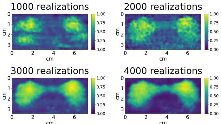
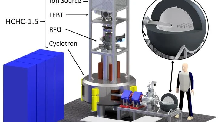
Treating cancer with a proton beam is a bit like archery: you need to hit the bull's-eye without hitting anything else. Protons release their energy at the end of their path, so healthy tissues are largely spared. But to make the shot safe, every treatment plan is checked in advance — measuring the dose the tumor will get and what the surrounding organs will receive.
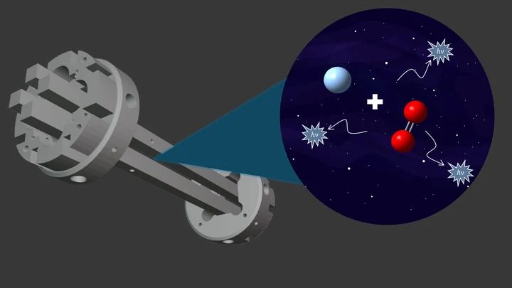
In the cold, sparse clouds of space, particles rarely meet, but sometimes an ion and a neutral molecule pass close enough to stick together. As they do, they emit light, like a muted flash carrying away excess energy. This process is called radiative association, and it is responsible for the formation of many molecules in interstellar space.