EEG Can't Tell Remission from Healthy express
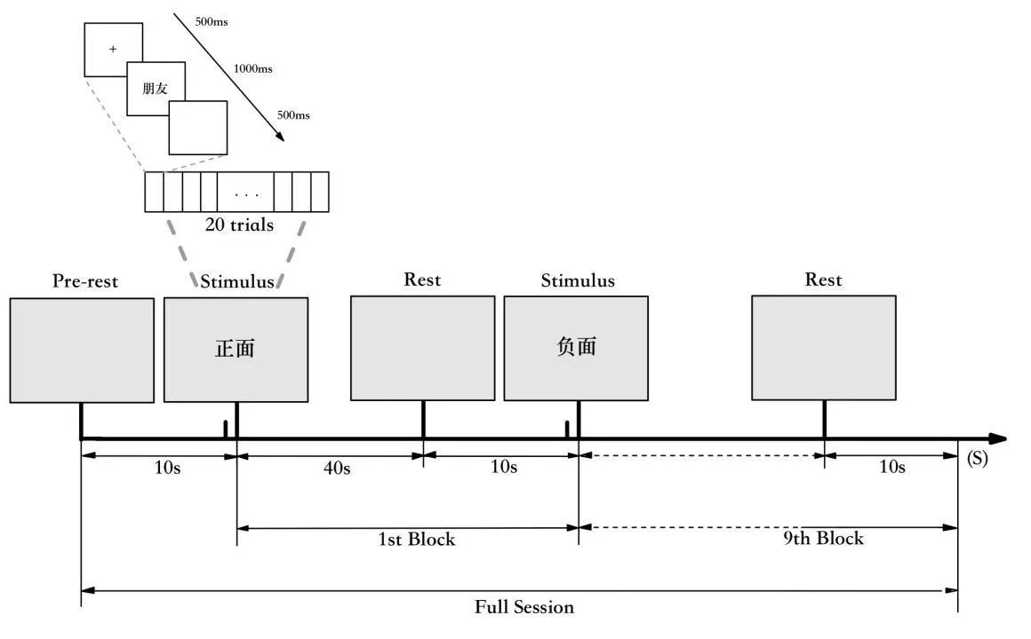
Depression is sometimes sought in the brain's electrical waves. The logic is simple: if a disease has stages (acute, maintenance, recovery) then the signals should change gradually, like volume when you turn a knob. Researchers took EEG recordings from 74 people: 24 in the acute phase, 23 in the maintenance phase, and 27 healthy. Then they checked whether classic wave measures really step up or down from illness to normal.
It turned out that almost no measure behaves so obediently. Each person's brain hums in its own way, and that hum drowns out the common pattern. The same number can appear in both a healthy person and a patient in remission. Group averages blur the picture rather than clarify it.
So, finding a biomarker for depression requires a different approach. Instead of comparing whole groups, track one person over time, like a specific patient, not an averaged portrait. Or build algorithms that learn from individual differences rather than group averages.
The volume knob turned out to be broken, but perhaps each device has its own scale.
Original abstract
as written by the authors arXiv:2603.03864
While EEG features differentiate Major Depressive Disorder (MDD) from healthy controls (HC), their clinical utility as biomarkers depends on a monotonic trajectory across the disease spectrum, from the acute (AC) phase to the maintenance (MA) phase and finally to the healthy baseline. However, the progression of the MA phase remains poorly understood in traditional marker analysis. Analyzing EEG data from 74 individuals (24 AC, 23 MA, and 27 HC), this study provides a comprehensive evaluation of classic ERP and resting-state indices across AC, MA, and HC groups. Our results demonstrate that almost no conventional metrics strictly satisfy the criterion of monotonic progression, likely due to profound inter-individual heterogeneity. These findings highlight the inherent limitations of group-level feature extraction and provide critical insights for developing future paradigms and algorithms to identify neurobiological markers with genuine clinical utility.
In healthy people, an EEG can change more from a cup of coffee or lack of sleep than in patients between phases of illness.