A microelectrode array (MEA) is a device consisting of hundreds or thousands of microscopicelectrodes arranged in a grid, designed to record or stimulate electrical activity from neurons, cardiac cells, or other excitable tissues. Unlike a single electrode, which captures the activity of one neuron, an MEA captures the simultaneous firing patterns of many cells, revealing how networks process information. MEAs are used both in vitro (on cultured cells) and in vivo (implanted in living brains), and they have become a cornerstone of modern neuroscience and bioengineering.
History
The first planar microelectrode arrays were developed in the early 1970s by C.A. Thomas and colleagues, building on earlier work with single microelectrodes. The technology matured through the 1990s with the advent of CMOS fabrication, allowing thousands of electrodes on a single chip.
How it works
Each electrode in the array is a tiny conductive contact, typically made of gold, platinum, or indium tin oxide, with a diameter of 10–30 micrometers. When a neuron fires, it produces a local voltage change that the electrode picks up; the signal is then amplified, digitized, and recorded. The spacing between electrodes is chosen to match the density of the cells under study — close enough to capture individual neurons, but far enough to avoid excessive crosstalk. Advanced MEAs can also stimulate cells by passing current through selected electrodes, enabling closed-loop experiments where activity is both read and written.
In practice
MEAs are used in drug screening for cardiac toxicity, in brain-machine interfaces for prosthetic control, and in basic research on learning, memory, and neural network dynamics.
Fun fact
Modern high-density MEAs can record from more than 20,000 neurons simultaneously — a number that exceeds the total number of neurons in the brain of a fruit fly.
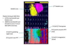
A radio telescope with thousands of antennas is like a giant ear that hears the whole sky at once. But an ear cannot draw a picture: signals from every pair of antennas must be multiplied, and in an array of ten thousand elements there are already fifty million pairs. A classical correlator does exactly that, and its complexity grows as the square of the number of antennas.
Blood vessel cells were seeded onto a transparent film just 700 nanometers thick — a hundred times thinner than a human hair. Flat electrodes are hidden beneath the film. They send a weak alternating current and listen as the resistance changes.
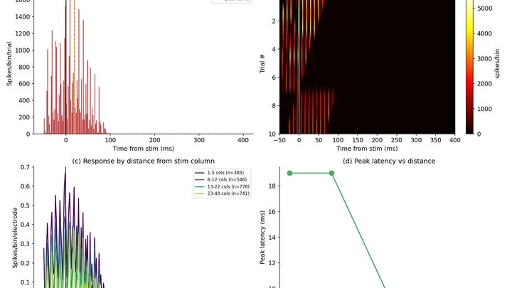
A stadium packed with fans hears the announcer’s voice for the first time, and instantly the whole place erupts. The message sounds again and again. Only a handful of people in nearby rows respond. That’s roughly how neural networks behave in brain organoids — tiny lab-grown replicas of the brain.
A thin sandwich made of two different materials. You run an electric current through it, and the top slice magnetizes in one direction, the bottom in the opposite. This isn't a magic trick; it's the layer Edelstein effect, just described by physicists.
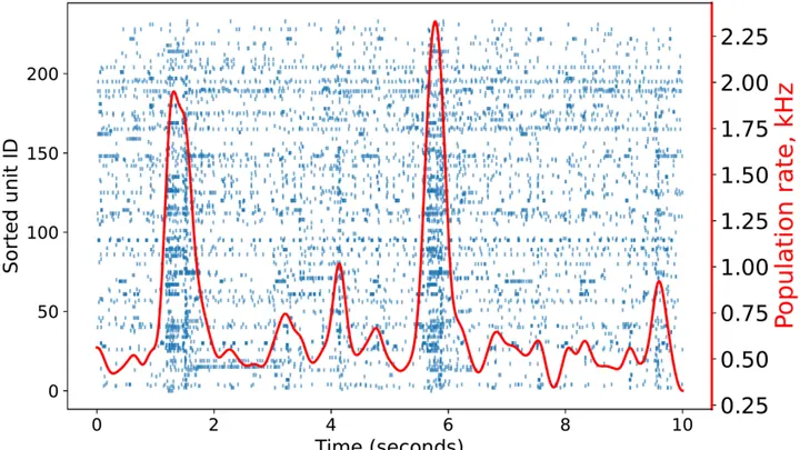
A city at night: lights on in some windows, dark in others. If you connect with lines the windows that light up simultaneously, you get a web of connections. Researchers studied such a web in tiny brain organoids — living clusters of nerve cells grown in the lab.
A base station emits radio waves. Without a special nozzle, the antenna acts like a garden hose opened full blast: water flies everywhere, but only a little reaches the flower beds. Researchers created a system with two neural networks.
A neuron is like a complex musical instrument. To understand how it responds to a touch, you used to have to connect to each string directly — a delicate, slow procedure that only works for individual cells.
The heart sometimes loses its rhythm, like an orchestra where the violins suddenly start playing each on their own. Cardiologists in such cases look for the "out-of-tune" areas by reading the heart's electrical signals and precisely ablating them with a thin probe — a catheter.
In absolute silence, you need to hear a feather drop. But someone keeps rustling a plastic bag. For the space-based LISA observatory, that 'rustling' will come from charged particles in space.
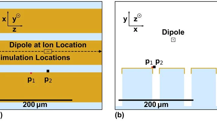
A quantum computer is like an orchestra where each ion is a musician and laser pulses are the conductor's baton. But if someone in the audience rustles, the delicate melody of quantum states goes off key.
A neuron resembles an electrical circuit with batteries and variable resistors. The resistors are played by ion channels — protein pores in the membrane that open and close, letting charged particles through. Whether a nerve impulse fires depends on how fast and at what voltage these pores operate.
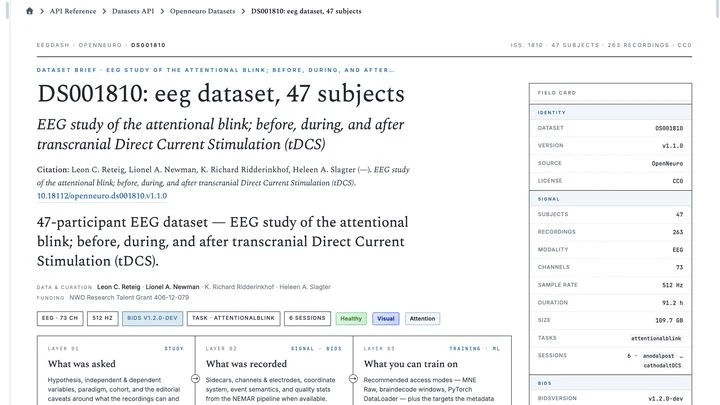
The open archives OpenNeuro and NEMAR hold 791 recordings of the brain's electrical activity from nearly forty thousand people. In total, that's 86,051 hours of signals — roughly ten years of continuous listening. Previously, to take even one such recording off the shelf, you had to write thousands of lines of code: download, unzip, fix format errors, and slice it into chunks of the desired length.
When the conductor leaves the orchestra, the musicians keep playing from memory for a while. But without a unified rhythm, each one starts to speed up or slow down on their own, and the harmonious symphony turns into disjointed noise. According to new research, the brain and heart behave much the same way in the last hours of a person's life.
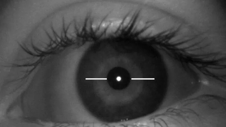
A flashlight shines into the eye, and the retina responds with electrical impulses, like an echo, but not of sound, of light. This echo can be recorded, and it reveals not only how the eye is working but also how the brain is. After all, the retina is a piece of the brain that has been pushed out to the periphery.
Biologists often tag cells with glowing molecules — it's like searching for a dim light in the smoke. The light quickly fades, and the smoke creates interference.
On the surface of a thin film of bismuth iron oxide, you can see neat stripes, like furrows in a plowed field. What lies beneath them has remained a mystery until now.
A cell is like a house with many doors. Some doors open only for certain guests, others let anyone through. To understand how these doors work, scientists build models of them. The new model is a glass slide with 52 miniature wells. Each well is covered with an ultrathin lipid film, like a jar with a lid. A protein that forms a channel through the film was inserted into it.
A colony of cells is like a city that grows from its outskirts: new houses appear where there's more space. Inside, it's crowded, and construction stalls. Similarly, cells divide more often at the edge, where neighbors squeeze them less. Scientists modeled this without any chemical commands — only mechanics.
In the model, each cell is a ball that presses on others.
An ordinary sheet of paper looks white because it scatters all light rays chaotically. But a butterfly's wing shimmers blue and green without a drop of paint: its surface is covered with microscopic grooves that dampen some light waves and amplify others. Engineers have long dreamed of replicating this trick: creating a material that colors itself simply by trapping light in the right places.
Depression is sometimes sought in the brain's electrical waves. The logic is simple: if a disease has stages (acute, maintenance, recovery) then the signals should change gradually, like volume when you turn a knob. Researchers took EEG recordings from 74 people: 24 in the acute phase, 23 in the maintenance phase, and 27 healthy.
Alzheimer's disease is like a tangled ball of yarn: thousands of genes are intertwined, and it's hard to tell which ones snap first. Previously, scientists looked for important genes by simply counting how many connections each node in the network had. But that approach is like judging a person by the number of acquaintances they have — the most sociable isn't always the most influential.
Ultracold molecules are a promising material for quantum computing, but assembling them into a neat array, like eggs in a carton, is no easy feat. A method has been proposed to load molecules one by one into optical tweezers or lattices directly from a gas. The molecules immediately end up in the calmest state of motion.
An electrocardiogram is like a long strip of paper on which the heart leaves its signature. A doctor studies it to catch rhythm disturbances, but when the recording lasts for days, the strip stretches for miles. The eye tires, attention wanders, and rare but dangerous irregularities can slip by.
AI-HEART works like a tireless assistant that scans the entire strip without a break.
Every thought leaves a trace — a faint electrical blip on the scalp. A device with dozens of wires, resembling a swim cap, catches these traces and turns them into wavy lines. From those lines, you can guess what a person feels even if they stay silent. In a new study, three computer programs competed to see which could read such lines most accurately and determine whether the mood was bad, neutral, or good.
A heart cell in a Petri dish contracts, and at that moment, an electrical wave sweeps across its surface. Previously, to catch that wave, you had to poke the cell with an ultra-fine electrode needle. Now, you just look at it under a microscope.
The method is called electrochromic recording.
A graphene transistor works like a microscopic switch: a molecule lands on its surface, and the current changes. But a bare graphene surface catches nothing. You need a "bait"—antibodies that recognize only viral proteins.
Silence in the operating room. The surgeon slowly lowers a needle thinner than a human hair into the patient's brain. At its tip is not a point but a whole palisade of nine hundred sixty microscopic microphones. Each one listens to its own tiny patch of gray matter.
Primary human cells, taken straight from the body, are the most honest material for studying diseases and testing drugs. But getting foreign genetic material into them is tricky: cells don't like intruders. Standard methods either damage the cell or work unevenly, especially when the sample is heterogeneous — which it almost always is.
The new platform tackles this with a vortex flow.
After an injection under the skin, a drug doesn't just spread out like ink in water. Antibody molecules carry an electric charge, and that charge makes them interact with surrounding tissues — much like a magnet attracts or repels iron filings.