A Scanner That Sees Through Tumors express
A standard PET scanner is like a camera with a narrow lens: to capture the whole body, you have to move it back and forth and take many shots. The new IMAS tomograph is a wide-angle camera that covers 71 centimeters of the body at once. The patient lies still, and the machine collects an image from head to thighs in a single pass.
Inside the scanner are rings of translucent crystals that catch flashes of light from a radioactive tracer injected into the blood. Each flash is a signal from a positron decay, and the more precisely the device determines where and when it occurred, the sharper the image. IMAS is the first to combine two techniques: measuring the time-of-flight of gamma photons and measuring the depth at which they are absorbed in the crystal. Previously, these capabilities were only available separately.
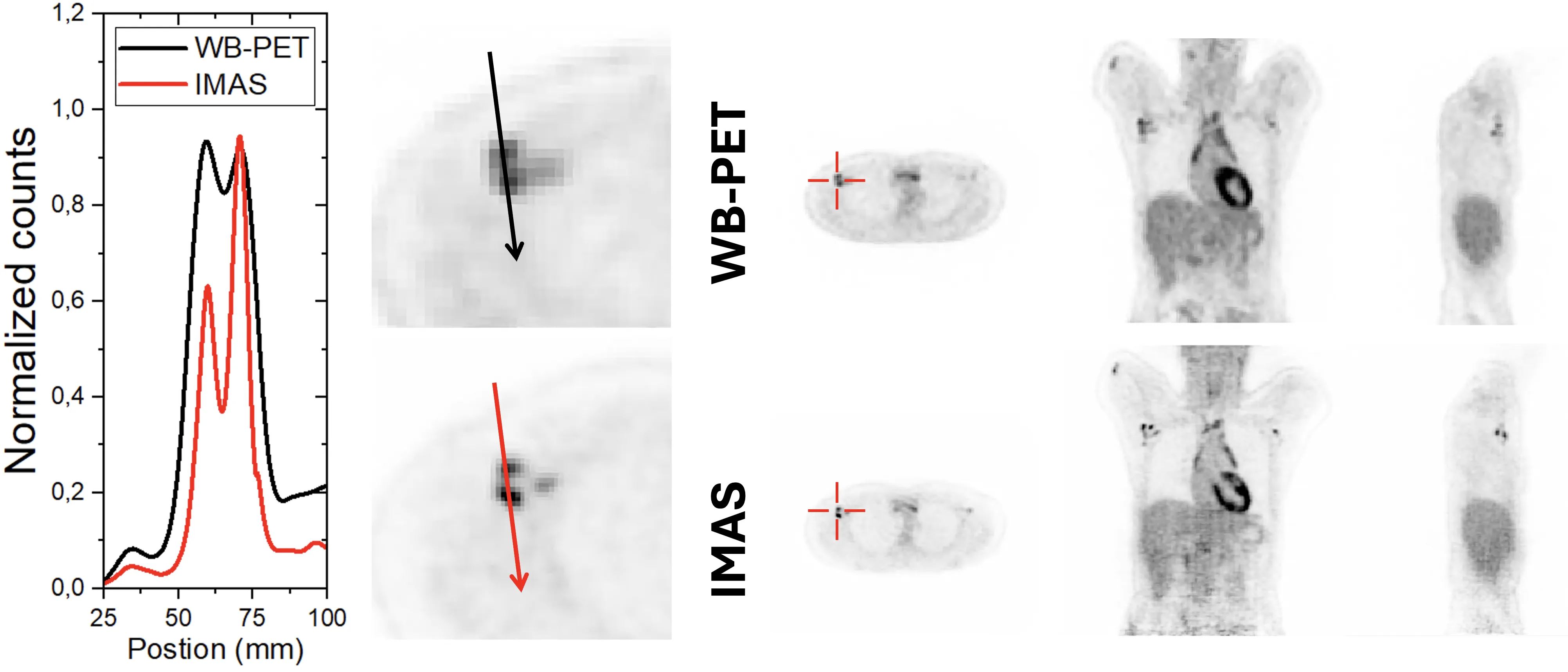
Initial tests on patients showed that the device sees tumors better than a commercial PET/CT scanner. However, the data acquisition speed is still below the design value — due to a bottleneck in transferring information to the computer. Engineers already know what to fix: replace the communication link, and the scanner will operate at full capacity.
Original abstract
as written by the authors arXiv:2603.09898
This work summarizes the design, construction, initial performance evaluation and pilot clinical results of the IMAS system, a long axial field of view (FOV), also known as total-body (TB-), positron emission tomography (PET) prototype scanner. This PET enables for the first time in TB-PET imaging, simultaneously time-of-flight (TOF) and depth-of-interaction (DOI) capabilities. The IMAS detector block is based on LYSO semi monolithic scintillators, with individual slab sizes of 3 mm x 25 mm x 20 mm each. Arrays of 1x8 slabs are coupled to 8x8 Silicon Photomultiplier arrays. A proprietary readout reduces the 64 signals to only 16 outputs, preserving both 3D photon impact positioning and timing accuracy. IMAS has a total of 30,720 channels. PETsys electronics is used for data acquisition. The IMAS geometry is based on 5 rings of 10 cm each, with a 5 cm gap between them. It defines an axial FOV of 71 cm with a bore aperture of 82 cm. We report in this work the pilot tests of the system performance and the first clinical results. We found that the system spatial resolution remained below 4 mm across the entire FOV, even at the off-radial position of 30 cm. A coincidence time resolution with a small size 22Na source of 560 ps FWHM was measured. A sensitivity of 56.54 cps/kBq is in good agreement with previous simulation studies; however, the noise equivalent count rates performance (79 kcps at 3.26 kBq/mL) was significantly lower than expected, likely due to a data transfer bottleneck between the system and the acquisition workstation. Finally, a comparison of one of the imaged patients with a commercial TOF PET/CT scanner is also provided, pinpointing an improved tumor identification for IMAS, and the advantages of TOF and especially DOI capabilities.
IMAS detectors are assembled from crystals that look like translucent bricks measuring 3×25×20 mm. Each brick acts like a miniature lens that not only captures light but also reveals the depth from which it came.