A tomograph is an imaging device that creates cross-sectional images of the body, allowing doctors to see inside without surgery. The term comes from the Greek 'tomos' (slice) and 'graph' (drawing). Unlike a standard X-ray, which superimposes all structures into a single flat image, a tomograph captures data from multiple angles and reconstructs it into detailed 2D slices or 3D volumes. This technology is foundational to modern medical diagnostics, enabling detection of tumors, internal injuries, and vascular abnormalities with remarkable precision.
History
The first clinically useful CT scanner was developed by Godfrey Hounsfield and Allan Cormack, who shared the 1979 Nobel Prize in Physiology or Medicine. Hounsfield built the prototype in 1971 at EMI Laboratories in the UK, and the first patient brain scan was performed in 1972. MRI followed, with the first human images produced in the late 1970s by Raymond Damadian and Paul Lauterbur.
How it works
Most clinical tomographs use X-rays (computed tomography, CT) or magnetic fields (magnetic resonance imaging, MRI). In CT, an X-ray tube rotates around the patient, emitting a fan-shaped beam; detectors on the opposite side measure attenuation. A computer algorithm, typically filtered back projection or iterative reconstruction, processes thousands of these measurements to build a matrix of tissue densities. In MRI, strong magnetic fields align hydrogen nuclei, and radiofrequency pulses perturb them; the emitted signals vary by tissue type, yielding high-contrast soft-tissue images. Both methods produce data that is essentially a numerical map, which the computer renders as grayscale images.
The first CT scanner took about 4 minutes to acquire a single slice and required the patient's head to be immersed in a water-filled rubber bag to reduce artifacts; modern scanners capture hundreds of slices in under a second.
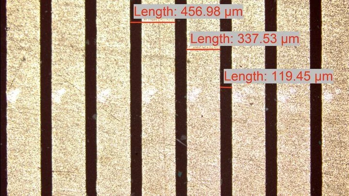
A radiologist looks at an abdominal scan and sees not just organs but their spatial relationships: which kidney is larger, where the tumor lies relative to a vessel. It's like reading a map, where it's not enough to know where a city usually stands; you also need to measure the distance between two points on the ground.
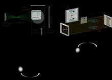
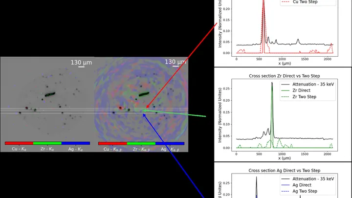
In a dark room, a sunbeam passes through a pinprick in cardboard, and an upside-down image of the street appears on the wall. A pinhole camera needs no lenses: the tiny hole itself selects the rays coming from each point of the scene. Scientists at the ESRF applied this principle not to light but to X-rays, and made the camera see not shape but chemical composition.
A doctor looks at a screen and sees two images of the same part of the body. One shows where blood is rich in oxygen, the other shows how fast it rushes through the finest vessels. Previously, these images were made by different devices, and combining them was difficult. Now engineers have built a device that takes both images simultaneously.
Two scans of the same brain taken at different times are like photographs of the same house before and after renovation. To see what changed, you overlay them and look at the differences. But an MRI scanner is not a perfect camera: it adds its own distortions, as if the photo were taken through slightly warped glass.
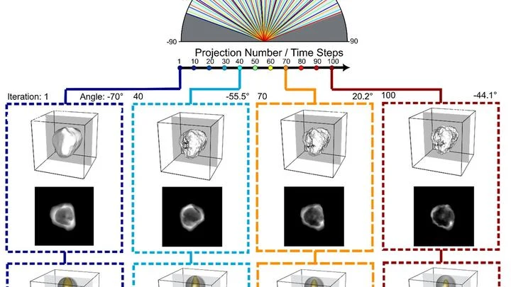
The data from a single CT scan is comparable to several feature-length films. TCAM-Diff bypasses this bottleneck: the program compresses the 3D scene into three shadow projections — like a sculptor reconstructing a solid statue from silhouettes on three walls.
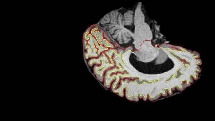
Medical scans are maps of tissue density. The brightness of each pixel reveals what’s there, much like photometry measures light intensity. But conventional algorithms analyze pixels in isolation, producing jagged organ boundaries.
A new approach compels the program to prioritize smoothness.
Tomographs don't produce ready-made pictures. They collect numerous 'shadows' — projections of innards from different angles or using various signals — and mathematically reconstruct a 3D picture from them.
Every second, about a hundred invisible particles, muons, pass through your body. They are born high in the sky when cosmic rays collide with air atoms, raining down on Earth. These particles are so penetrating that they can pass through a multi-story building, but they slightly change direction when they encounter dense material, such as lead or uranium.
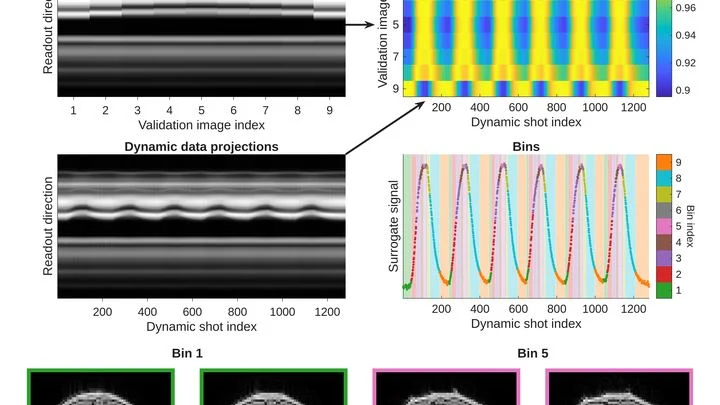
A photographer shoots a runner with a long exposure and gets a blurry silhouette. That's roughly how a standard MRI scanner "sees" moving organs: the heart contracts, the lungs breathe, and the image smears. To avoid this, patients are asked to freeze and hold their breath. But for a child or someone short of breath, that's nearly impossible.
The method called GRASP tackles the problem differently.
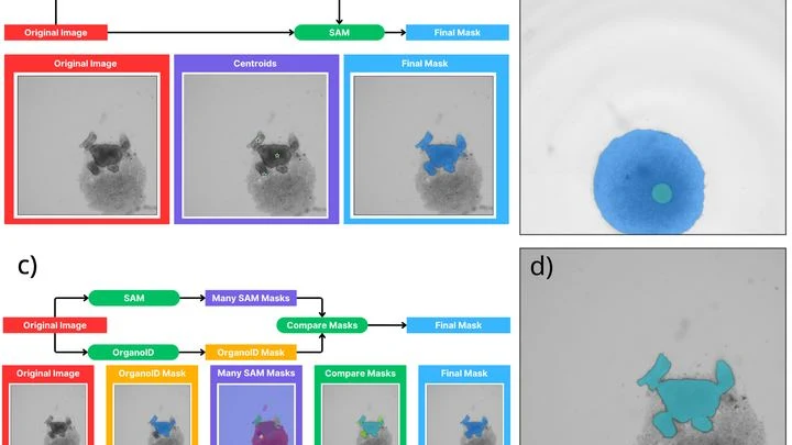
A biologist looks through a microscope at dozens of tiny cell spheres — spheroids. From these, miniature organ-like structures will later grow. The task is similar to a baker's job of checking whether the dough has risen evenly in each mold. Only here, instead of molds, there's a nutrient medium, and instead of dough, there are living cells.
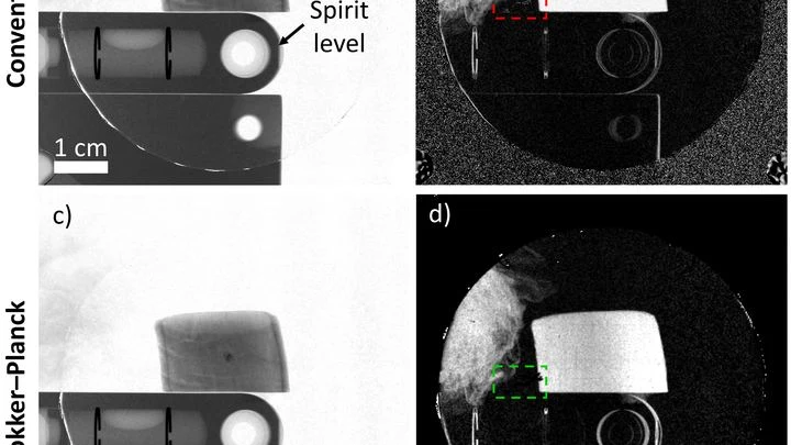
An X-ray machine with gratings takes not one but three pictures at once: a regular image, a map of beam deflections invisible to the eye, and an image of tiny structures that scatter light. Together, they help spot, for example, emphysema or pulmonary fibrosis. But there's a catch: to separate these three layers, you need a clever mathematical trick.
Two specialists look at the same breast image. One is an experienced doctor, the other is an artificial intelligence program. Sometimes their opinions match, sometimes they diverge. How often they agree determines how accurately they can together distinguish healthy tissue from a tumor.
Conventional X-ray fluorescence analysis works like a meticulous scribe: to learn the composition of a picture, it walks over every square centimeter and logs whether lead or copper is present. For a flat sheet, that's tolerable, but for a three-dimensional object, it's torture.
A mole on your skin looks like a mosaic of colored pieces. Each piece is a small patch of the image where color and texture barely change. Such pieces are called superpixels. If you connect them with lines, you get a network that looks like a subway map: stations are the pieces, and lines are the connections between them. The weight of each line shows how strongly two pieces are related.
A tiny gold star melts under the beam of an electron microscope, its points slowly rounding off. In the past, such a process could only be seen in snapshots taken with long pauses, and the probing beam itself would alter the sample.
A finger is illuminated with a laser beam, and some of the light returns after traveling inside. That's how Raman spectroscopy works: molecules in bone and skin alter the color of the reflected beam differently, leaving their chemical fingerprints. But skin, muscle, and fat are noisy: their signal drowns out the bone's, like a crowd's chatter overpowering a single singer.
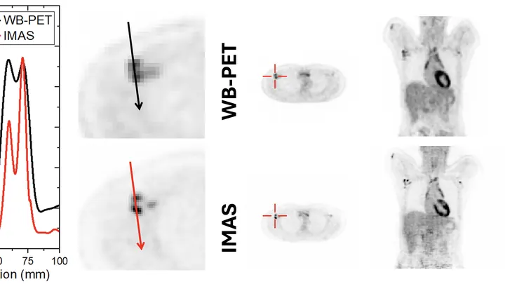
A standard PET scanner is like a camera with a narrow lens: to capture the whole body, you have to move it back and forth and take many shots. The new IMAS tomograph is a wide-angle camera that covers 71 centimeters of the body at once. The patient lies still, and the machine collects an image from head to thighs in a single pass.
A fat droplet inside a living cell is like a bobber in rough water. It doesn't move in a straight line but in jerks, sometimes pausing, sometimes speeding up. Previously, such movements could only be seen in a single plane, like watching the bobber from above without knowing its depth.
Your back hurts, and the doctor asks you to bend forward and backward to see how your vertebrae move. Usually, this means a series of X-rays: they show motion, but only in a plane, like a shadow on a wall. And volumetric methods like CT scans can't keep up with movement.
Engineers proposed a different approach: a robot sweeps an ultrasound probe over your back while you bend.
A photographer snaps a runner: to avoid a blurry shot, a short shutter speed is needed. An MRI scanner is like that photographer, but its 'shutter speed' is long — by the time it captures, the muscle has already changed. That's why dynamic MRI (a series of volumetric frames in motion) is a tough challenge: data are scarce, and motion doesn't wait.
Radioactive tracers are like a courier with a beacon: you inject it into the body, and it highlights the target organ on the scan. But this courier comes with strict rules: you can't send it often, it quickly 'gets tired,' and it costs a lot. Doctors have long dreamed of a way to see an organ without radiation.
Light is a great tool for studying tissues, but it fades fast deep inside the body. Within a few millimeters, photons scatter so much that you can't make out anything. Scientists came up with a workaround: instead of shining light from outside, deliver a tiny light source right inside. The challenge is how to power that source without wires.
An X-ray image is like a photograph taken in the dark with a very short flash: anything that moves comes out blurred. Conventional X-ray detectors are large arrays of millions of pixels, and by the time they read out one frame, the next is already missed. For a spinning turbine or a beating heart, such sluggishness is a death sentence.
A team of researchers sidestepped this problem by ditching the array altogether.