A Scanner That Sees Through Tumors express
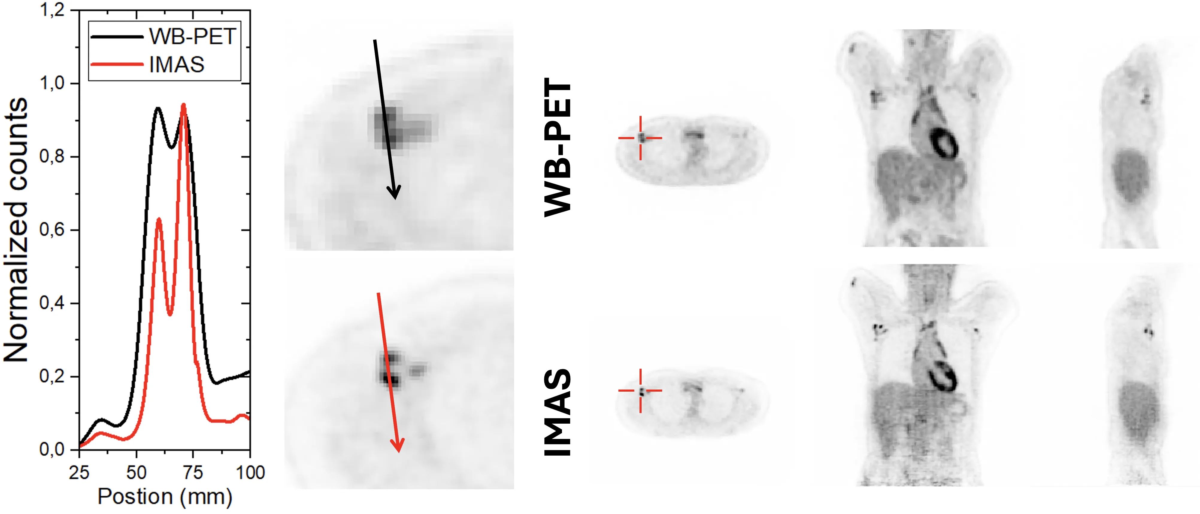
A standard PET scanner is like a camera with a narrow lens: to capture the whole body, you have to move it back and forth and take many shots. The new IMAS tomograph is a wide-angle camera that covers 71 centimeters of the body at once. The patient lies still, and the machine collects an image from head to thighs in a single pass.
Inside the scanner are rings of translucent crystals that catch flashes of light from a radioactive tracer injected into the blood. Each flash is a signal from a positron decay, and the more precisely the device determines where and when it occurred, the sharper the image. IMAS is the first to combine two techniques: measuring the time-of-flight of gamma photons and measuring the depth at which they are absorbed in the crystal. Previously, these capabilities were only available separately.
Initial tests on patients showed that the device sees tumors better than a commercial PET/CT scanner. However, the data acquisition speed is still below the design value — due to a bottleneck in transferring information to the computer. Engineers already know what to fix: replace the communication link, and the scanner will operate at full capacity.
IMAS detectors are assembled from crystals that look like translucent bricks measuring 3×25×20 mm. Each brick acts like a miniature lens that not only captures light but also reveals the depth from which it came.