Resolution is the ability of an imaging system to distinguish between two closely spaced objects or details. In physics and engineering, it is a fundamental limit that determines how fine a structure can be observed, whether in a microscope, telescope, camera, or medical scanner. Higher resolution means finer details can be seen, which is crucial for fields ranging from astronomy to materials science.
History
The concept of resolution limits was formalized by Lord Rayleigh in the 19th century, who defined a criterion for when two point sources are just barely distinguishable. This Rayleigh criterion remains a standard reference.
How it works
Resolution is limited by the wavelength of the radiation used and the numerical aperture of the optical system. For conventional optics, the diffraction limit sets a minimum angular separation below which two points blur into one. Techniques like using shorter wavelengths (e.g., X-rays instead of visible light) or computational methods (e.g., super-resolution) can push beyond this limit. In digital imaging, resolution also depends on pixel size and sampling density.
In practice
Resolution is critical in X-ray microscopy, medical imaging (CT, MRI), satellite photography, and optical lithography for microchip manufacturing.
Fun fact
The Hubble Space Telescope, with a mirror of 2.4 meters, can resolve details as small as about 0.05 arcseconds — equivalent to reading a newspaper from about a kilometer away.
A camera looks at two distant sparks. They're so close together that they merge into a single fuzzy blob. It seems impossible to tell them apart — the classic limit of optical microscopy has been reached.
But quantum mechanics hints that the light field still holds a secret about where each spark sits.
An array of radio telescopes is like a camera missing several lenses. Ever since Karl Jansky picked up the radio hiss of the Milky Way, astronomers have been trying to piece together a whole picture from fragmentary signals. But the antennas are spaced apart, and the mathematically “developed” image — the dirty map — ends up smeared.
Imagine photographing a statue in dim light. You face a dilemma: take one bright shot or a series of dim frames from different angles. In the first case, quality is limited by photon noise, the graininess that arises from a lack of photons, while in the second, details overlap, making it harder to reconstruct the 3D structure.
A surgeon removes a breast tumor, but how do you know no cancer cells are left at the edge? Right now, the tissue goes to a lab, and the answer comes back days later. If the margin is not clean, a second surgery is needed. The new method promises to give the answer right in the operating room, while the patient is still on the table.
A conventional light microscope hits a fundamental limit: it cannot distinguish two points closer than 200 nanometers — roughly the thickness of a soap bubble divided by a hundred. To see deeper, scientists have made molecules in the sample blink or fade, but these tricks are slow and harmful to living cells.
An X-ray image usually brings to mind a bulky machine in a hospital. But such a picture can be taken by a device the size of a palm, assembled from hobbyist electronics. The authors took a Raspberry Pi mini-computer, a 12.3-megapixel camera, and a screen that glows under X-rays.
An X-ray image of a battery looks like a photo taken through frosted glass: fine details blur into a gray smudge. The culprit is the detector itself, which cannot distinguish features smaller than its pixel size. Engineers proposed getting around this barrier by illuminating the sample not with a uniform beam but with a striped pattern.
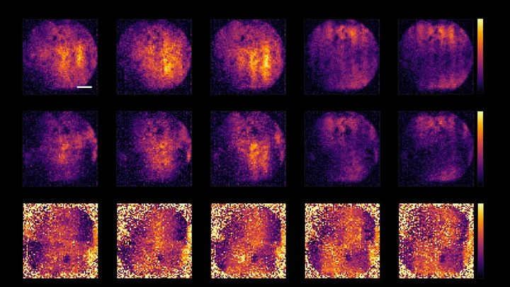
Our body's tissue is like a city: different neighborhoods are home to different cells, each with its own job. To understand how this city is organized, biologists take images showing which genes are active at each spot. But sharp images are expensive, and cheap ones come out blurry, like a photo of a city from high above, where individual buildings are hard to make out.
In the dark, a tiny spark flashes. A gamma ray born inside a patient's body strikes a crystal and knocks out a shower of light. The detector's job is to figure out exactly where that happened. The more precise the answer, the sharper the image on the tomograph screen.
The new detector resembles a honeycomb, but not a flat one — the cells narrow toward the bottom.