Positron emission tomography (PET) is a medical imaging technique that produces three-dimensional images of functional processes in the body, such as glucose metabolism or blood flow. It works by detecting pairs of gamma rays emitted indirectly by a radioactive tracer that has been injected into the patient. PET is widely used in oncology, neurology, and cardiology to diagnose diseases, assess treatment response, and guide surgical planning.
History
The underlying physics was established in the 1950s, and the first PET scanner was developed in the early 1970s by researchers including Michael Phelps and Edward Hoffman at Washington University. The technique became clinically widespread in the 1980s and 1990s.
How it works
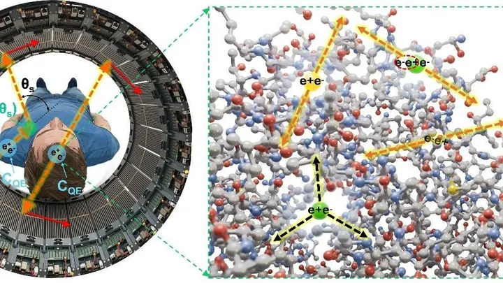
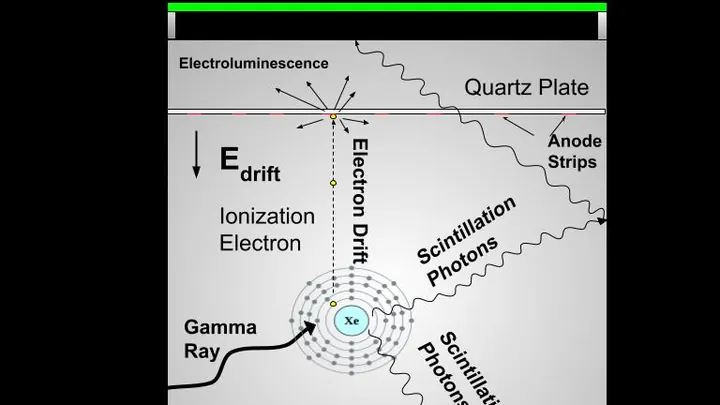
A radioactive isotope attached to a biologically active molecule — typically a glucose analog like FDG — is introduced into the body. As the isotope decays, it emits a positron, which travels a short distance before annihilating with an electron. The annihilation produces two gamma-ray photons traveling in exactly opposite directions. A ring of detectors around the patient records these simultaneous photon pairs, and computer algorithms reconstruct the spatial distribution of the tracer, revealing areas of high metabolic activity.
In practice
PET is essential for cancer staging and monitoring, detecting metastases, evaluating brain disorders like Alzheimer's disease, and assessing myocardial viability in cardiology.
Fun fact
The two gamma rays from a positron annihilation are emitted back-to-back, which is why PET detectors are arranged in a ring — they must catch both photons to locate the event.
Light falling on matter usually scatters off it or heats it up. But when there are many photons and they are very energetic, two quanta of light can collide and vanish, leaving two particles in their place: an electron and its antimatter twin, the positron. This process, predicted back in 1934, has never been reproduced in the lab with real photons.
The electron beam of a microscope usually flies through emptiness, like a courier on an empty highway. Any air molecule is a bump that makes it bounce and lose direction. So before, to examine a sample, you had to freeze it, dry it, or coat it with metal—and still you could only look in a vacuum.
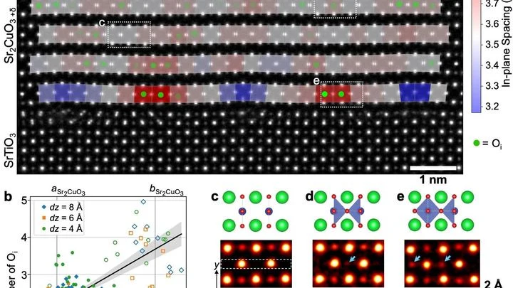
A superconductor is a material that conducts electricity without loss, like water flowing through a perfectly smooth pipe. In copper-based superconductors, tiny oxygen additions act like a volume knob: a bit more and the material starts superconducting; a bit less and it stops. But the oxygen atoms themselves are so small that seeing them directly was impossible.
When a laser beam hits a gas, it tears electrons from atoms and accelerates them to nearly the speed of light. As these electrons fly through the plasma, they emit X-rays, like a glowing trail from a fast particle. In the new work, physicists made a single laser pulse create not one but two such trails in succession.
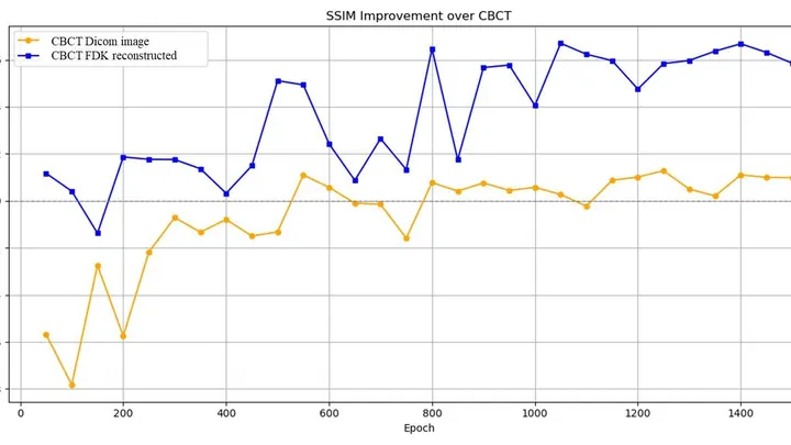
A low-dose X-ray image looks like a view through a fogged-up window: outlines are there, but details drown in a hazy veil. Doctors need such images during radiation therapy — to aim the beam precisely at the tumor and spare healthy tissue. But saving on radiation comes at the cost of noise, streaks, and artifacts that obscure what matters.
Photographing an exoplanet orbiting another star is like trying to spot a firefly in front of a spotlight. Earth's atmosphere is constantly churning, which makes stars appear as dancing speckles through a telescope.
Tomographs don't produce ready-made pictures. They collect numerous 'shadows' — projections of innards from different angles or using various signals — and mathematically reconstruct a 3D picture from them.
Each positron annihilation is like the birth of a pair of dancers, connected by an invisible thread of polarization. Two gamma quanta fly apart, but their states remain entangled: measuring one instantly determines the other — as if one partner senses every movement of the other, no matter the distance.
Isolating the signal from a single pair of supermassive black holes is like hearing a dancer's footsteps amid the roar of a hurricane. Somewhere in space, these monsters spin in a slow gravitational tango, distorting spacetime itself.
A radiologist stares at gray-white smudges on the screen — dozens of body slices where every suspicious spot must be found. Manual segmentation of one patient takes half an hour, and a large clinic sees hundreds of such patients. Fatigue and varying experience inevitably introduce variability: two specialists will outline the same lesion slightly differently.
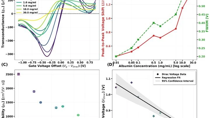
In a drop of blood float millions of albumin molecules — the main transport protein. Its level drops in kidney and liver diseases, so doctors need a fast and accurate way to measure it. Engineers have proposed a detector the size of a dust particle: a graphene transistor that works directly in liquid.
Graphene is a sheet of carbon one atom thick.
The heart sometimes loses its rhythm, like an orchestra where the violins suddenly start playing each on their own. Cardiologists in such cases look for the "out-of-tune" areas by reading the heart's electrical signals and precisely ablating them with a thin probe — a catheter.
Ever since Paul Dirac theoretically predicted antimatter, physicists have been hunting for the tiniest differences between particles and their antiparticles.
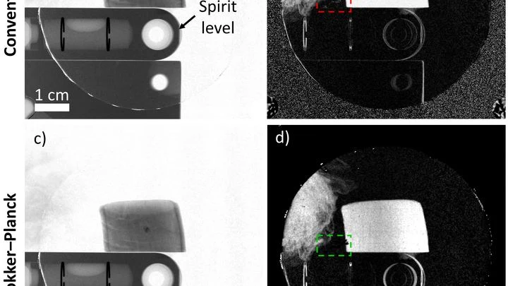
An X-ray machine with gratings takes not one but three pictures at once: a regular image, a map of beam deflections invisible to the eye, and an image of tiny structures that scatter light. Together, they help spot, for example, emphysema or pulmonary fibrosis. But there's a catch: to separate these three layers, you need a clever mathematical trick.
You taste a fruit smoothie through two different straws. One lets through only the big chunks, the other only the liquid. Individually, the sensations are misleading, but together they give you the key: here, it seems, is banana, and here, strawberry.
When matter is heated to millions of degrees, it turns into plasma — a mix of free electrons and ions, like a boiling soup of charged particles. You can't measure the temperature of such a 'soup' with an ordinary thermometer, but you can shine X-rays through it.
Two photons fly out from a single point in a patient's chest. They are twins: alter one, and you instantly know about the other. This connection is called quantum entanglement. In a PET scan, these pairs are born inside the body when an injected radioactive substance meets electrons in the tissues.
The degree of entanglement is not constant.
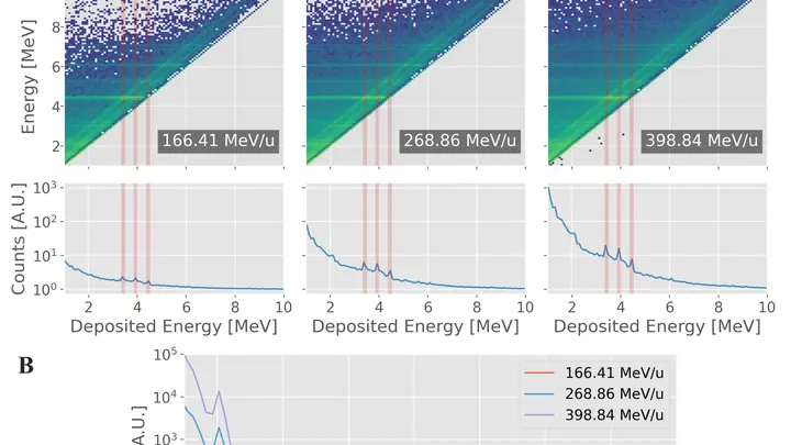
When a carbon ion beam enters the body, it doesn't release energy all at once but rather at the end of its path, like a runner who gives their all on the final stretch. Doctors need to know this finish point to avoid damaging healthy tissue. But how can you look inside the body without making an incision?
Your finger is a living sensor: beneath the skin, arteries pulse in time with your heart. Traditional blood pressure cuffs squeeze your arm and give a single number, but this ring uses eight electrodes to pass a weak current through the tissue and map blood flow.
Inside every cell, there are tiny power stations — mitochondria. They burn nutrients and store energy. Two helper molecules, NADH and FAD, oversee this process. Previously, they were detected by their faint glow, but it was dim, and signals from different molecules overlapped.
The new microscope works differently.
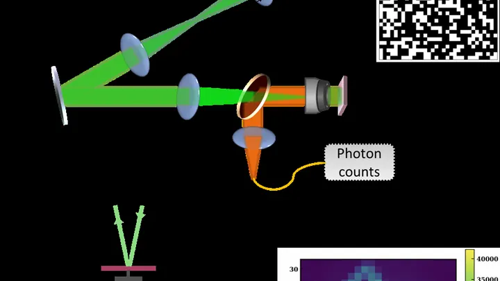
Hunting for a single glowing spot in a dark sample is like trying to find a needle in a haystack: the microscope beam crawls line by line, wasting time and light. Physicists replaced this slow sweep with random light patterns. The sample is illuminated with a broad field, but each time with a new pattern of bright and dark patches, as if random shadows from a grid were projected onto it.
A PET scanner catches flashes of light that occur when a radioactive tracer introduced into the body meets electrons in tissues. Usually, the device merely notes where a flash occurred. But between the birth of the tracer and the flash, there is a pause — tiny, in billionths of a second.
The radioactive isotope terbium-149 acts like a tiny homing bomb: it latches onto cancer cells and detonates with alpha particles that destroy the tumor from within, barely touching healthy tissue. But this bomb has a quirk: it also glows with positrons, and that glow can be captured by a special camera.
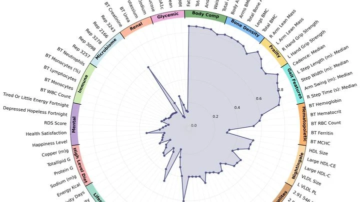
Gait is not just a way to get from point A to point B. Each step, arm swing, and torso tilt is like a signature the body leaves in space. Doctors have long noticed that disease alters gait, but they considered it merely a symptom of a specific problem: if you limp, your leg hurts. Now it turns out that gait is a whole book about health, and you can read it with an ordinary camera.
The camera that hunts for tumors works like a highly sensitive camera: it catches the faint glow from a substance that accumulates in diseased cells. In conventional machines, the light is born in a crystal — a solid piece that resembles glass. But a crystal doesn't always convey exactly where the light flashed, so the image comes out slightly blurry.
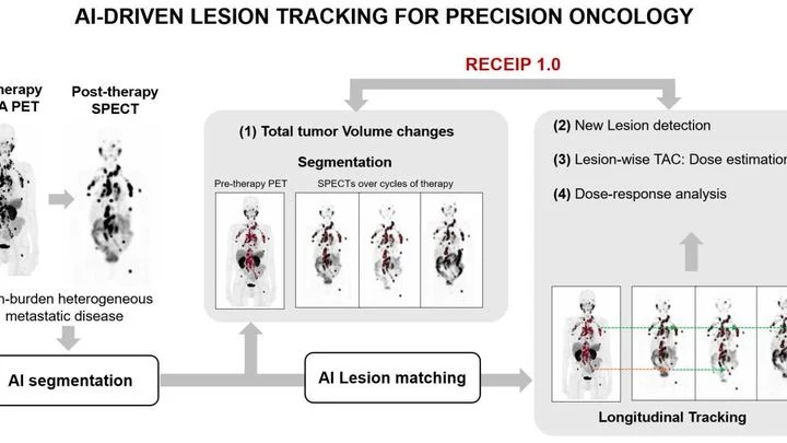
When treating prostate cancer, doctors often see not one tumor but many lesions in bones and lymph nodes. Each lesion can respond to therapy differently: one shrinks, another stays the same, a third grows. Previously, the disease was assessed as a whole, like judging the weather by the average temperature over a year.
PET/CT is like a map where cancer cells light up as bright spots. But what if the map is blank and the disease is still hiding somewhere? In 132 men with recurrent prostate cancer, doctors found no glowing lesions, but they decided to study the texture of the scan itself, like an experienced tracker noticing bent grass where an animal has passed.
A beam of carbon ions strikes the tumor precisely, like a surgeon who can only see the skin. Inside the body, the beam stops, and exactly where used to be a mystery. Now, helium is mixed into the same beam: it passes through the body like an X-ray, and its trail shows where the carbon went.
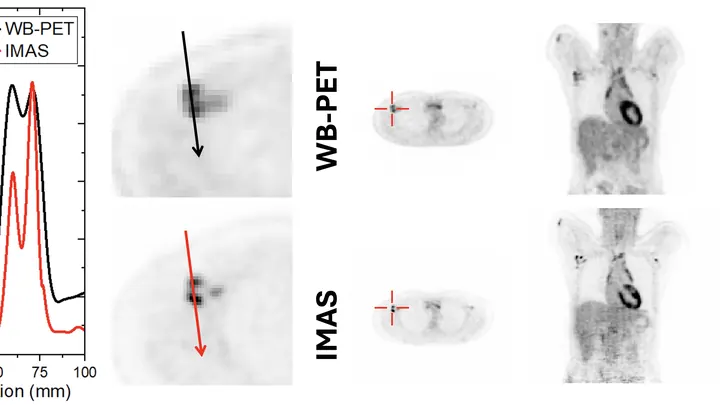
A standard PET scanner is like a camera with a narrow lens: to capture the whole body, you have to move it back and forth and take many shots. The new IMAS tomograph is a wide-angle camera that covers 71 centimeters of the body at once. The patient lies still, and the machine collects an image from head to thighs in a single pass.
A scanning tunneling microscope probes a molecule with an ultra-fine tip and maps its relief. But the image is flat, while the molecule is three-dimensional. How do you guess a person's pose from their shadow on a wall?
A lightning bolt strikes an anthill. Some ants die instantly, some freeze, and a few days later the survivors start rebuilding the tunnels. That's roughly how a tumor behaves after electroporation — a method where short electric pulses are passed through tissue to poke holes in cancer cell membranes and kill them.
A fat droplet inside a living cell is like a bobber in rough water. It doesn't move in a straight line but in jerks, sometimes pausing, sometimes speeding up. Previously, such movements could only be seen in a single plane, like watching the bobber from above without knowing its depth.
When treating epilepsy, doctors adjust the drug dose almost blindly. Too little, and seizures continue; too much, and side effects appear. Usually, a patient's blood is sent to a lab, and the result comes back a day or two later. Now, the same test can be done on a tiny silicon chip.
The sensor works like a highly sensitive nose.
A fingernail-sized piece of ceramic is squeezed in a vise, and its internal structure rearranges like clay under your fingers. Inside, orderly rows of tiny domains line up where they were once randomly oriented. After this 'massage,' the crystal lets light through almost like glass and, at the same time, generates a strong electric charge when bent or pressed.
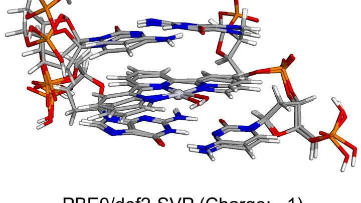
Standard chemotherapy hits the whole body like carpet bombing: both cancerous and healthy cells suffer. Scientists are hunting for more precise weapons — molecules that light up when they slip into a cancer cell's DNA, revealing its location. Such glowing probes could help doctors spot tumors early and even destroy them with pinpoint accuracy, leaving neighboring tissue untouched.
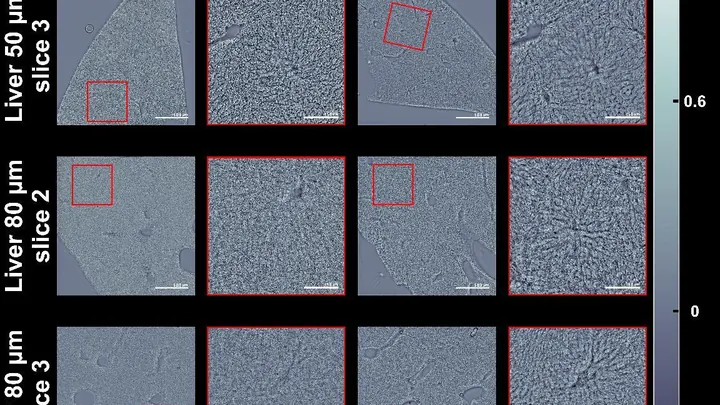
A thick slice of tissue under a microscope looks like fogged glass: light scatters and details are lost. Usually, to see cells, you stain them or cut them into very thin slices. But there's a simpler way — make the slice itself transparent, as if you've wiped the glass clean.
Hidden in the nucleus of a gold atom lies a potential future cancer treatment. When a gold target is struck by a gamma ray with an energy of about 30 megaelectronvolts, one of the nuclei loses a proton and a neutron, transforming into a rare platinum isotope — platinum-195m.
Radioactive tracers are like a courier with a beacon: you inject it into the body, and it highlights the target organ on the scan. But this courier comes with strict rules: you can't send it often, it quickly 'gets tired,' and it costs a lot. Doctors have long dreamed of a way to see an organ without radiation.
Imagine a vast field of fireflies, each flashing alone rather than in a swarm: that's what the new automated setup is hunting for. It sweeps a laser across the sample like a flashlight and records the color and brightness of the glow at every spot.
A firefighter with a thermal imager can see the source of a fire through smoke. But how do you see a radiation source that has no smell, no glow, and no heat? Gamma cameras solve this problem: they capture gamma rays emitted by radioactive materials and overlay their 'trace' onto a regular photograph. The result is an image showing that the source is, say, behind the far door.