Your Smartphone Instead of an X-Ray: How a Photo of Your Body Predicts Health express
The scale tells you your weight, but it's silent about where those pounds sit — in muscle or belly fat. And it's the fat tucked deep inside, around your organs, that's most often linked to heart disease and diabetes. Previously, you could only see it with an expensive scanner at a clinic. Now, a smartphone is all you need.
Scientists trained the program on tens of thousands of photos of people whose body composition had been precisely measured. Then they tested it on new volunteers: the program estimated body fat percentage with an error of only about two percent. For comparison, the usual body mass index (weight divided by height squared) often gets it wrong because it can't tell muscle from fat.
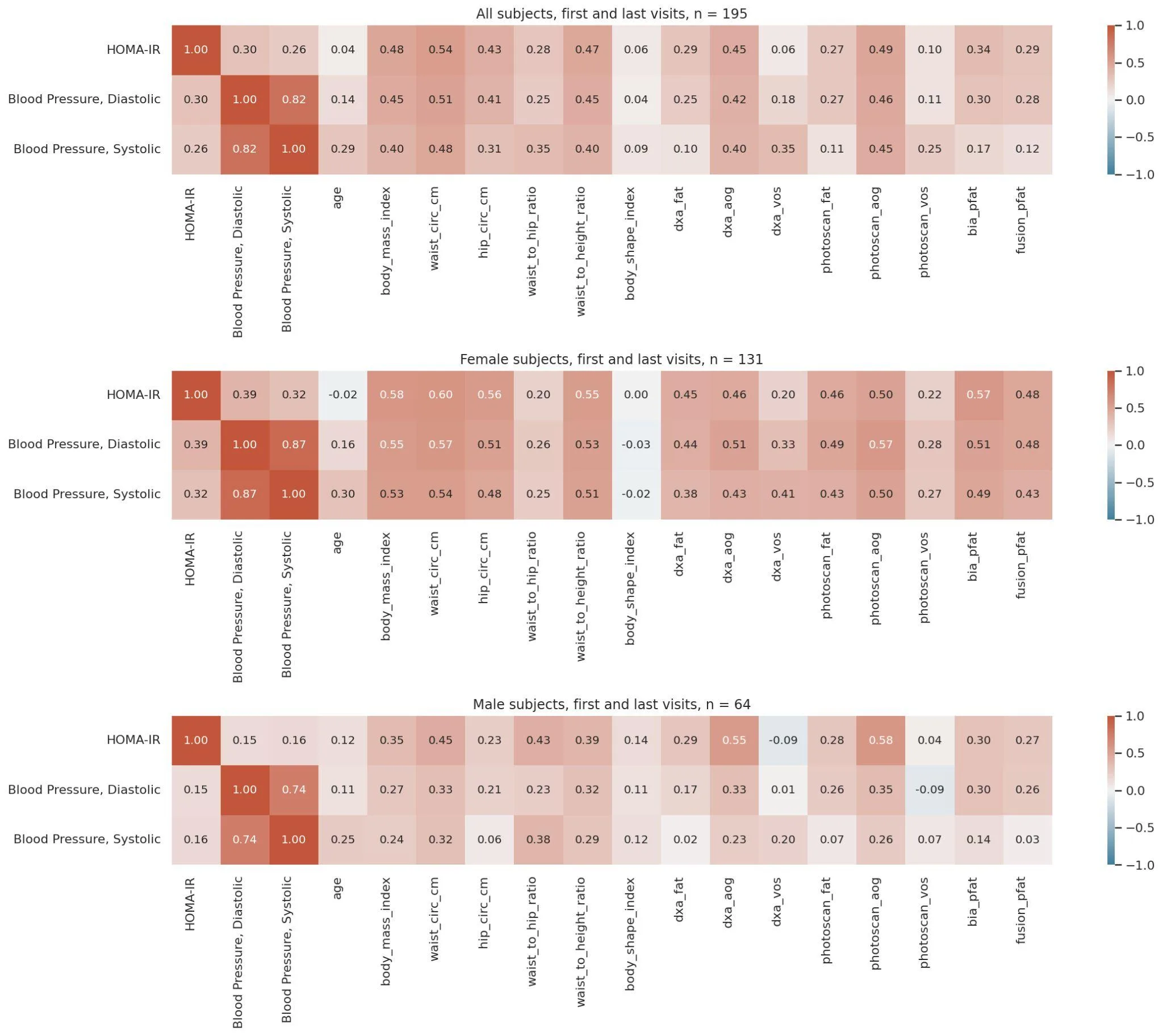
When they added the photo-based measurements to data on age, sex, and body mass index, the accuracy of predicting insulin resistance, the first step toward diabetes, improved noticeably. And it performed almost as well as using an expensive scanner. That means a simple snapshot could serve as an early warning sign for a doctor.
This approach won't replace a doctor, but it could make body composition screening accessible to millions. Instead of waiting in line for a complex exam, a photo can help catch the risk in time.
Original abstract
as written by the authors arXiv:2603.27017
Body Mass Index (BMI) is a widely accessible but imprecise proxy of cardiometabolic health. While assessing true body composition is superior, gold-standard methods like Dual-Energy X-ray Absorptiometry (DXA) are not scalable. We address this gap by developing and validating "PhotoScan," a method to estimate body composition from smartphone imagery. We pretrained a deep learning model on UK Biobank participants (N=35,323) and fine-tuned on a newly recruited clinical cohort (PhotoBIA cohort, N=677) with diverse ethnicity, age, and body fat distribution, achieving high accuracy against DXA for total body fat percentage (BF%, MAE = 2.15%), Android-to-Gynoid fat ratio (A/G, MAE = 0.11), and visceral-to-subcutaneous fat area ratio (V/S, MAE = 0.09). Generalizability of the model was demonstrated on an independent metabolic health study cohort (MetabolicMosaic cohort, N=132 participants), achieving MAEs of 2.13% for BF%, 0.09 for A/G, and 0.09 for V/S. We then evaluated the clinical utility of these metrics in the MetabolicMosaic cohort by predicting insulin resistance (IR). Adding PhotoScan-derived body composition metrics to baseline demographics model (Age, Sex, BMI) significantly improved insulin resistance classification (Area Under the Receiver Operating Characteristic Curve "AUROC" 76.0% vs 69.2%, DeLong test p=0.002, Net Reclassification Index "NRI" 0.593). Crucially, this accessible smartphone method achieved performance nearly equivalent to adding clinical-grade DXA data to baseline demographics model (AUROC 77.3% vs 69.2%, DeLong test p=0.004, NRI 0.748). These findings demonstrate that smartphone-based phenotyping captures clinically meaningful risk signals missed by BMI and anthropometrics, offering a scalable alternative to DXA for cardiometabolic risk stratification.
The body mass index was invented nearly 200 years ago for statistics, not for assessing an individual's health. It doesn't account for how much muscle you have versus fat.