Medical image analysis is the field where artificial intelligence and neural networks are trained to interpret medical images such as MRI scans, CT scans, and optical spectroscopy data. The goal is to detect pathologies like cancer, predict patient outcomes, and assist clinicians in making decisions. Instead of relying solely on a radiologist's visual inspection, AI models can identify subtle patterns in images that may not be obvious to the human eye, such as early signs of tumors or changes in tissue structure.
History
The use of computers in medical imaging dates back to the 1960s, but modern deep learning approaches emerged after 2012, when convolutional networks achieved breakthroughs in general image recognition. Since then, the field has rapidly expanded with applications in radiology, pathology, and oncology.
How it works
Neural networks, particularly convolutional neural networks (CNNs), are trained on large datasets of labeled medical images. The network learns to map image features—like edges, textures, and shapes—to diagnostic labels. For example, in MRI scans of brain tumors, the network can learn to distinguish between healthy tissue and tumor regions by analyzing pixel intensity patterns and spatial relationships. Once trained, the model can process a new image and output a probability score for the presence of a disease or a predicted outcome, such as response to chemotherapy.
In practice
AI-based medical image analysis is used in hospitals to screen for cancers, segment tumors, and predict treatment responses, such as in MRI scans for glioblastoma or prostate cancer.
Fun fact
Some AI models can predict a patient's brain age from an MRI scan, and a significant gap between predicted and chronological age may indicate neurodegenerative disease.
Imagine trying to figure out what happened in a room from the sound that lingers after a gunshot. The shot is gone, but the echo still bounces off the walls. That's roughly how a neuropathologist views a brain tissue section where the tumor itself is absent: the biopsy only captured healthy tissue that reacted to the disease.
At night on a volcano summit, six telescopes stare at the same speck of light. Each catches its own copy of the wave, and if you combine their signals, you get an image that no single instrument could produce alone. But there's a catch: light captured by different stations cannot simply be added like photographs.
A radiologist looks at an abdominal scan and sees not just organs but their spatial relationships: which kidney is larger, where the tumor lies relative to a vessel. It's like reading a map, where it's not enough to know where a city usually stands; you also need to measure the distance between two points on the ground.
A forensic examiner arrives at a crime scene and lifts fingerprints. Epigenetic clocks work the same way: from chemical marks on DNA they determine how much a cell has aged. A tumor leaves its own "fingerprints," and almost always they point to decrepitude.
The researchers took 5528 samples from the TCGA database, which holds DNA methylation data for eight cancer types.
In the orchestra of our immunity there are three conductors, and each has a score of their own. One tells the musicians to play softer when a threat appears in the hall, another does the opposite and cranks up the noise, and the third conducts in such a way that the hall fills with sound, though nobody can say why.
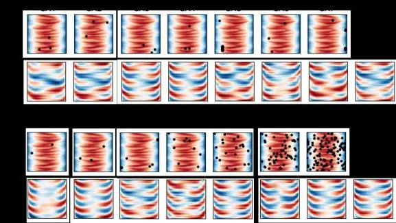
Every heartbeat sends a wave through the cerebral arteries that not only pushes blood but also briefly inflates the vessel. The arterial wall, like a tightly stretched rubber tube, passes that motion to the neighboring brain tissue, and the brain pulses ever so slightly in time with the heartbeat.
A telescope stares at a star for many nights in a row. Somewhere in the stream of light hides a tiny dip in brightness — an Earth-sized planet briefly blocking part of the star. Such a dip lasts a few hours and repeats once a year or less. Catching it is like hearing a single quiet click in a noisy hall.
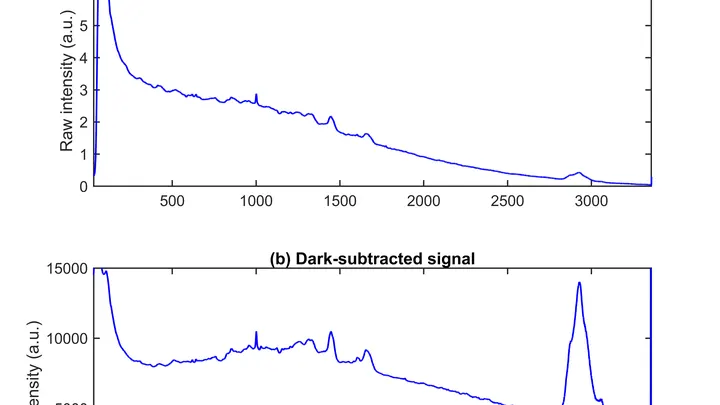
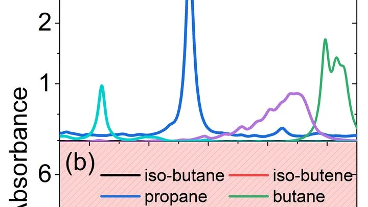
The instrument shines an infrared beam at a sample, and molecules respond with their own absorption pattern, like bells of different tones. This pattern, the spectrum, holds information about the chemical composition. But to read it, a specialist usually consults a library of reference records, which is slow and not always possible.
A doctor spends years learning to recognize diseases from brain scans. First, they excel at spotting signs of aging, then they master tumors, and suddenly they start confusing what they knew before. Many neural networks behave much the same way: new knowledge overwrites the old.
Predicting the yield of a chemical reaction is a bit like trying to guess the harvest knowing only the soil composition. Traditionally, chemists have relied on long columns of numbers, quantum descriptors, that describe electron energies but say nothing about molecular shape.
The Sun continuously scribbles magnetic fields across its surface. These patterns are not mere ripples but a handwriting in which nature has encoded warnings of flares. A flare is an explosion that releases the energy of billions of nuclear bombs in minutes, ejecting streams of plasma and radiation into space.
Ordinary AI looks at medical images like puzzles made of unfamiliar pieces: it considers any two unmatched pictures completely different. It's like trying to tell twins apart without knowing they're related. In medicine, however, patients with the same condition look nearly identical on MRI, and the algorithm mistakes 'relatives'.
Genes are like houses in a city: in some, life bustles; in others, silence reigns. This 'urban' genetic landscape was analyzed in a thousand women with breast cancer.
Communicating with a satellite is like trying to hear a whisper in a storm: the signal drowns in noise. Engineers have created an AI algorithm that runs directly on the satellite and, with minimal power consumption, detects distortions more accurately than previous methods.
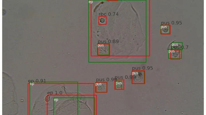
A drop of urine under a microscope isn't a flat puddle but a multi-layered volume. Like a stack of transparent pages: if you focus on one, the neighboring ones blur. The microscope's light only shows one depth layer sharply. To see all the cells, a lab technician had to take a series of snapshots, manually adjusting focus — tedious and slow.
Assessing image quality is a job for a meticulous inspector with a toolbox. The IQA-T1 neural network acts exactly like that: it doesn't just stare at the picture, but applies either a spectroscope or a light meter.
Neutron stars are the last clumps of visible matter before collapsing into a black hole. In their interiors, matter is squeezed to trillion-fold densities. Here, nuclear forces and gravity weave into an exotic substance inaccessible to any earthly particle accelerator.
A cook who learns to cook from hand-drawn pictures risks ruining every dish. The same trap awaits computer vision: models need thousands of labeled images, but collecting them manually is expensive and tedious. Synthetic frames solve the problem, but only if they look real.
Prostate MRI scans often look like photos taken through a dirty window: noise, blurred edges, curvature from field imperfections. Scientists checked what happens if you 'wash' that window: remove the excess, correct the distortions, and accurately align different types of scans.
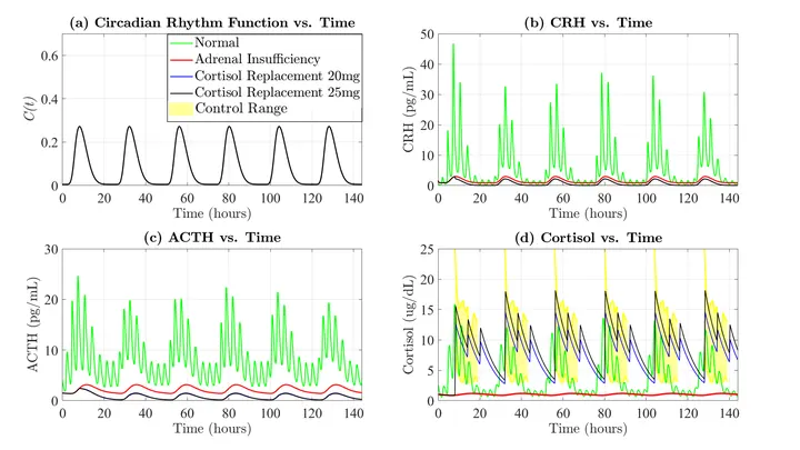
Inside each of us, invisible clocks are ticking. They control the release of cortisol — the hormone that helps us wake up in the morning, cope with anxiety, and keep our immune system on its toes. In adrenal insufficiency, these clocks break down: the body can't produce the right amount of cortisol on its own.
Dozens of copper coils in an MRI scanner pick up the faint response from tissues, like microphones trying to catch a whisper in a noisy hall. Each microphone drags a thick bundle of wires behind it. The wires press on each other, create electrical interference, heat up, and add risk for the patient.
Inside a fusion reactor, plasma rages — a gas heated to millions of degrees. There are no direct windows, and cameras and sensors quickly fail. Engineers see only fragments of information: a few sensors at scattered points. It's like trying to grasp a symphony from the sound of three microphones in a vast hall.
The eye is inflamed, the cornea clouding. The doctor needs to quickly determine the culprit — bacteria or fungus. The treatment depends on it. A mistake can cost sight. But the classic lab answer takes days, and in remote areas it may never arrive.
Researchers from the US and India propose a different approach: three images from a slit lamp — a device found in nearly every eye clinic.
A radiologist stares at gray-white smudges on the screen — dozens of body slices where every suspicious spot must be found. Manual segmentation of one patient takes half an hour, and a large clinic sees hundreds of such patients. Fatigue and varying experience inevitably introduce variability: two specialists will outline the same lesion slightly differently.
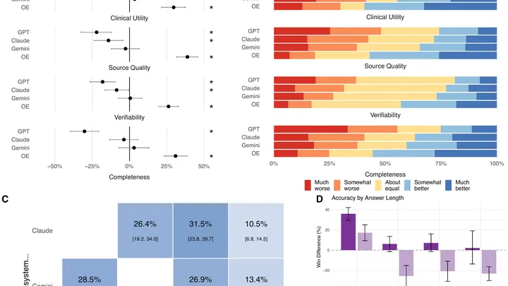
Every week, doctors ask AI assistants millions of questions at the bedside. But these tools are usually tested on exam-style problems, not on what clinicians actually ask in practice. A team of researchers set out to fix that.
They collected 620 real questions from physicians across 30 specialties — from cardiology to oncology.
Geophysics has always sought a language to converse with the depths. Well drilling is a crude alphabet, laboratory core tests are slow decoding. A new approach turns humming porosity into a pure note.
Solar flares are powerful explosions on the Sun that can knock out satellites and blackout cities. Predicting them is complicated by the tangled magnetic fields in sunspots.
X-ray detectors work like honeycombs: particles flash here and there in hexagonal cells. Conventional algorithms stretch these combs onto a square grid, creating ghostly polarization artifacts. Scientists used a neural network that lives by the rules of the hive — it processes the hexagonal structure directly, preserving the natural pattern.
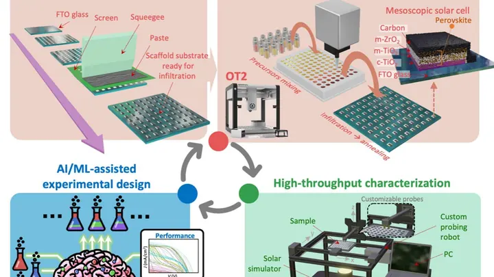
Perovskite crystals harvest sunlight almost as well as silicon, but cost pennies. Their trouble is an inborn fragility: from warmth and ordinary water in the air, they “get sick.” Inside, disorder builds up, and the shiny plate turns cloudy, losing its abilities within a couple of days.
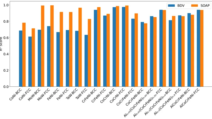
Creating an alloy from several metals is like cooking a soup from mysterious ingredients: guessing the final texture without trial and error is nearly impossible. Conventional models often get it wrong when there are more than three different kinds of atoms.
The scene is lit by spotlights, but the most interesting action happens in the shadows — where molecules invisible to the eye are hiding. The new camera picks up the heat of these "invisible" objects and turns it into ordinary light.
A proofreader checks a manuscript before it goes to print. They don't know every possible mistake, but they know exactly what flawless text looks like. Spotting a strange symbol or a blurred word, they immediately circle it in red. That's roughly how the new program for checking medical images works.
Your arm lifts a cup from the shelf — a familiar motion you don't notice until muscles tire. Then the trajectory changes: the elbow shifts slightly, the hand trembles. A doctor usually catches these changes by hand, feeling the patient's muscles.
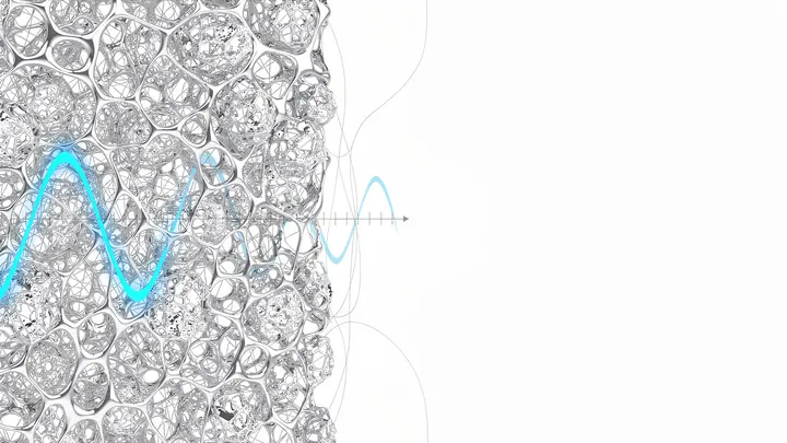
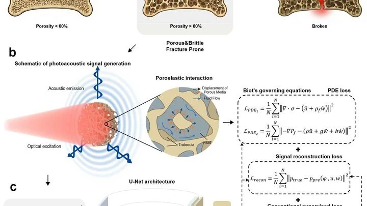
A laser beam strikes bone, and it responds with a nearly inaudible ultrasonic echo. Hidden in that sound is information about every microcrack and every fluid-filled pore. The trouble is that conventional computer programs don't understand this language: they see the signal but don't know the physics that produced it.
When you need to see inside the brain, you usually lie in a tight tube of an MRI scanner or get zapped with X-rays. Both methods are costly, bulky, and not always available.
A drop of blood from a vein falls under three beams, and each one drags its own clues into the light. The infrared beam makes molecules vibrate — from that trembling, you learn how much sugar and fat is in the sample. A green laser scatters off proteins and lipids, shifting color — revealing the remodeling of cell membranes.
An X-ray tomograph is like a carpenter who examines a log from all sides to see where the void is inside. It takes dozens of flat shadow images, rotating the source and detector around the object. Then a 'reconstruction' program glues these shadows into a 3D model. The quickest reconstructor is the FBP algorithm (filtered back projection).
A hundred people are talking at once in a large hall. You try to hear one, but voices blend, some whisper, and you don't even suspect the existence of others. That's roughly how a conventional laser gas analyzer feels when dozens of substances mix in a combustion chamber or a factory pipe.
The light bulb turns on only in the room where you need it. That's roughly how photoactivatable drugs work: they 'switch on' only where light is directed, leaving the rest of the body untouched. This is especially important in cancer treatment: conventional chemotherapy hits all dividing cells indiscriminately, causing severe side effects.
A neutron spectrometer is like a blind geologist who taps on rock and guesses what's inside by the sound. It sends a beam of neutrons into a crystal and watches how they scatter. From this pattern, you can deduce how atomic magnets, spins, oscillate. But where exactly to dig and how long to listen used to be decided by a human.
Now, the TAS-AI algorithm has taken the helm.