Photographic body analysis uses standard photographs or full-body images to extract health-related information. By analyzing subtle features like skin color, body shape, posture, or even the pattern of blood flow in the face, algorithms can estimate physiological parameters such as heart rate, blood pressure, or the risk of metabolic diseases. This approach is non-invasive, low-cost, and can be performed with a smartphone, making it accessible for remote monitoring and large-scale screening.
History
The idea of diagnosing from appearance dates back to ancient medicine, but the modern computational approach emerged in the 2010s with deep learning and the ubiquity of smartphone cameras.
How it works
The method relies on computer vision and machine learning. The algorithm detects key landmarks on the body, then analyzes pixel-level variations—for instance, the slight changes in skin color caused by blood pulsing under the skin (photoplethysmography). It can also measure body composition from silhouette analysis or detect jaundice from the yellowness of the sclera. The model is trained on large datasets where the same individuals have both photos and clinical measurements, allowing it to learn correlations between visual features and health outcomes.
In practice
It is used in telemedicine apps to monitor chronic conditions, in fitness trackers to estimate heart rate from the face, and in research to screen for diseases like anemia or diabetes from selfies.
Fun fact
Some algorithms can detect a person's age, gender, and even stress level from a single photo, with accuracy comparable to clinical measurements.
A stain is found at a crime scene. The expert takes a swab, and a few hours later the instrument outputs a list of proteins that made up the fluid. Previously, a rapid test could be wrong if multiple fluids were present: blood mixed with saliva, and the strip gave a false result.
A treasure map drawn by a neural network promises to show where the gold is buried. But if you take that map to another island, the marks may point to nothing. That is exactly what happened with the explanations from a model that estimates pulse from facial video.
The RhythmFormer neural network picks up subtle color changes in the skin caused by heartbeats.
A translator at the negotiating table doesn't listen to every word; they grasp the meaning of whole phrases. Electron microscopy poses a similar challenge: you need to figure out the crystal's thickness and its tilt in the beam, and all you have is a diffraction pattern, a mosaic of thousands of bright and dark spots.
Imagine trying to figure out what happened in a room from the sound that lingers after a gunshot. The shot is gone, but the echo still bounces off the walls. That's roughly how a neuropathologist views a brain tissue section where the tumor itself is absent: the biopsy only captured healthy tissue that reacted to the disease.
In 2023, in Belgium, 101 people aged 50 to 69 spent several days in a lab where their brains were scanned, their sleep was recorded, and they were asked to solve memory and attention tasks. That is how the COFITAGE dataset was born, describing each participant from five angles: molecular, structural, physiological, genetic and cognitive.
Every heartbeat sends a wave through the cerebral arteries that not only pushes blood but also briefly inflates the vessel. The arterial wall, like a tightly stretched rubber tube, passes that motion to the neighboring brain tissue, and the brain pulses ever so slightly in time with the heartbeat.
A doctor looks at a screen and sees two images of the same part of the body. One shows where blood is rich in oxygen, the other shows how fast it rushes through the finest vessels. Previously, these images were made by different devices, and combining them was difficult. Now engineers have built a device that takes both images simultaneously.
Light that never reaches the camera still leaves its mark on it. That's how the new imaging method works: you illuminate the object with one color, and the picture is assembled from another. Inside the crystal, light waves mix, and information about the invisible transfers to a visible beam.
In the experiment, they used a silver thiogallate crystal.
In a quiet room of a Budapest laboratory, a bracelet lies on the table, and instead of a strap, it has a thin conductive thread stretched in a zigzag. It doesn't heat up or glow, but when pressed against the wrist, it starts to listen. Each heartbeat sends a pressure wave through the radial artery, and the thread, deforming, changes its electrical resistance.
A drop of blood from a fingertip lands on the white membrane of a cartridge. Twenty minutes later, the phone shows whether Lyme disease is present or not. No test tube, no centrifuge, no waiting for days.
Usually, for such a test, blood is first spun in a centrifuge to separate the liquid part — the plasma.
A glucose meter that doesn't prick your finger but just looks at your wrist: that's the dream for millions of people with diabetes. It's the promise of algorithms that extract glucose levels from the ordinary pulse signal, the photoplethysmogram (the same one that measures heart rate in smartwatches).
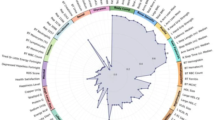
An X-ray machine takes a scan, and the doctor sees only numbers — bone density, fat percentage. But the image holds far more. A team of researchers created a model called LeDXA that reads these scans like a map of hidden bodily signals.
The model was trained on 11,540 scans from the Human Phenotype Project with no labels at all.
Genes are like houses in a city: in some, life bustles; in others, silence reigns. This 'urban' genetic landscape was analyzed in a thousand women with breast cancer.
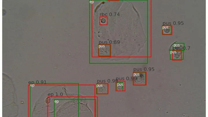
A drop of urine under a microscope isn't a flat puddle but a multi-layered volume. Like a stack of transparent pages: if you focus on one, the neighboring ones blur. The microscope's light only shows one depth layer sharply. To see all the cells, a lab technician had to take a series of snapshots, manually adjusting focus — tedious and slow.
Imagine a kinked garden hose: water meets resistance, and pressure drops. A similar process occurs in arteries when they lose their straight geometry. Vascular tortuosity is not just a cosmetic defect but a factor that disrupts hemodynamics and poses a threat of ischemic stroke.
Tomographs don't produce ready-made pictures. They collect numerous 'shadows' — projections of innards from different angles or using various signals — and mathematically reconstruct a 3D picture from them.
A radiologist stares at gray-white smudges on the screen — dozens of body slices where every suspicious spot must be found. Manual segmentation of one patient takes half an hour, and a large clinic sees hundreds of such patients. Fatigue and varying experience inevitably introduce variability: two specialists will outline the same lesion slightly differently.
The heart sometimes loses its rhythm, like an orchestra where the violins suddenly start playing each on their own. Cardiologists in such cases look for the "out-of-tune" areas by reading the heart's electrical signals and precisely ablating them with a thin probe — a catheter.
Diabetes is a constant reminder of needles. To check glycated hemoglobin (HbA1c — a blood protein that reflects average sugar over three months), you need regular blood draws from a vein. It's unpleasant, requires a lab, and takes time. And if you live far from a city, every test becomes a major expedition.
The new iGLU 5.0 device changes the game.
A proofreader checks a manuscript before it goes to print. They don't know every possible mistake, but they know exactly what flawless text looks like. Spotting a strange symbol or a blurred word, they immediately circle it in red. That's roughly how the new program for checking medical images works.
Your arm lifts a cup from the shelf — a familiar motion you don't notice until muscles tire. Then the trajectory changes: the elbow shifts slightly, the hand trembles. A doctor usually catches these changes by hand, feeling the patient's muscles.
When you need to see inside the brain, you usually lie in a tight tube of an MRI scanner or get zapped with X-rays. Both methods are costly, bulky, and not always available.
An X-ray tomograph is like a carpenter who examines a log from all sides to see where the void is inside. It takes dozens of flat shadow images, rotating the source and detector around the object. Then a 'reconstruction' program glues these shadows into a 3D model. The quickest reconstructor is the FBP algorithm (filtered back projection).
To peek inside a muscle, it is illuminated with radio waves in a strong magnetic field: that's how MRI works. Water in cells and water in blood respond differently, but a standard scanner doesn't tell them apart: it produces an averaged image where the signal from capillaries is superimposed on the signal from muscle fibers.
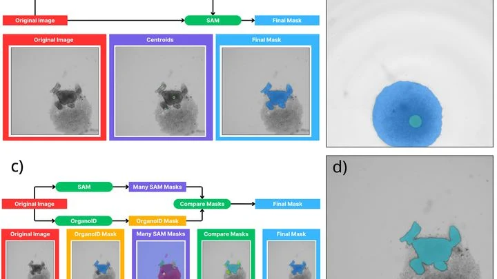
A biologist looks through a microscope at dozens of tiny cell spheres — spheroids. From these, miniature organ-like structures will later grow. The task is similar to a baker's job of checking whether the dough has risen evenly in each mold. Only here, instead of molds, there's a nutrient medium, and instead of dough, there are living cells.
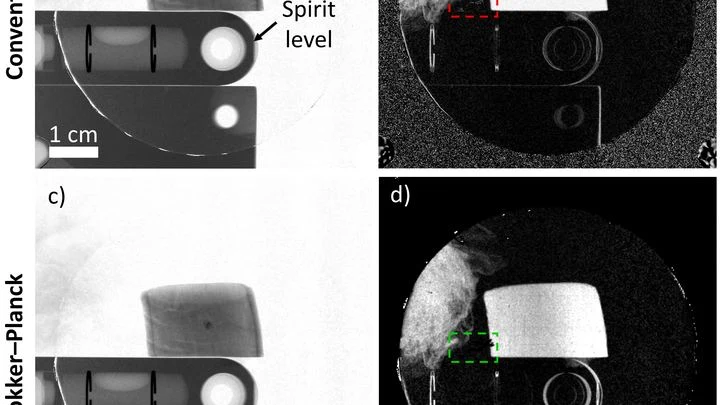
An X-ray machine with gratings takes not one but three pictures at once: a regular image, a map of beam deflections invisible to the eye, and an image of tiny structures that scatter light. Together, they help spot, for example, emphysema or pulmonary fibrosis. But there's a catch: to separate these three layers, you need a clever mathematical trick.
When a person breathes, air doesn't flow in a steady stream but in waves. Previously, doctors and scientists only looked at overall numbers: how much air entered per breath, what the peak flow was. But within a single breath, there are nuances — small variations that speak to the workings of nerves and muscles. The new approach breaks a breath into parts, much like a musician breaks a chord into individual notes.
An ordinary thread sewn into fabric can listen to the body. It feels the chest rising with each breath, a finger bending, and the pulse beating at the wrist. All this — without stiff patches or wires, just as part of your clothing.
The sensor is made from a flexible thread that changes resistance when stretched or pressed. It is woven into fabric in a triangular pattern, like a fine mesh.
Each heartbeat can be drawn as a point on a sheet of paper. Thousands of these points merge into a cloud. The Mapper algorithm turns this cloud into a network: it connects points that behave similarly and colors them according to blood flow strength. The result is not just a pile of numbers but a colored map showing which parts of the heart work in sync and which are out of step.
Your finger is a living sensor: beneath the skin, arteries pulse in time with your heart. Traditional blood pressure cuffs squeeze your arm and give a single number, but this ring uses eight electrodes to pass a weak current through the tissue and map blood flow.
Inside every cell, there are tiny power stations — mitochondria. They burn nutrients and store energy. Two helper molecules, NADH and FAD, oversee this process. Previously, they were detected by their faint glow, but it was dim, and signals from different molecules overlapped.
The new microscope works differently.
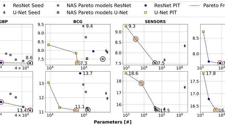
Smartwatches measure your pulse with light: an LED shines through the skin, and a sensor picks up the reflected signal. By analyzing the fluctuations in this signal, you can estimate blood pressure, but accurate calculation requires a powerful neural network — usually running in the cloud, so data leaves the device.
Brain aging is like the wear and tear on a bridge: cracks appear unnoticed, but if you reinforce the supports in time, the structure lasts longer. Three ordinary things turned out to be such supports — handgrip strength, the ability to move for a long time without breathlessness, and lung capacity.
A mole on your skin looks like a mosaic of colored pieces. Each piece is a small patch of the image where color and texture barely change. Such pieces are called superpixels. If you connect them with lines, you get a network that looks like a subway map: stations are the pieces, and lines are the connections between them. The weight of each line shows how strongly two pieces are related.
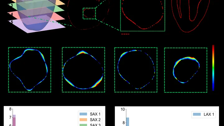
The heart is not a perfect sphere; its shape changes with every beat. To build an accurate 3D copy for a computer model, doctors typically take dozens of MRI slices. But that's like assembling a watermelon from thin slices: the fewer slices, the more guesswork.
A new approach teaches a neural network to understand the heart's shape from sparse contours.
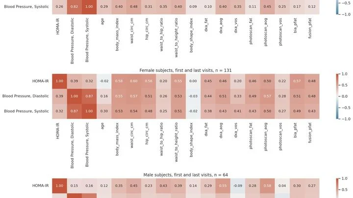
The scale tells you your weight, but it's silent about where those pounds sit — in muscle or belly fat. And it's the fat tucked deep inside, around your organs, that's most often linked to heart disease and diabetes. Previously, you could only see it with an expensive scanner at a clinic. Now, a smartphone is all you need.
Gait is not just a way to get from point A to point B. Each step, arm swing, and torso tilt is like a signature the body leaves in space. Doctors have long noticed that disease alters gait, but they considered it merely a symptom of a specific problem: if you limp, your leg hurts. Now it turns out that gait is a whole book about health, and you can read it with an ordinary camera.
In a hospital, blood from a finger prick usually goes to the lab, and the result comes back the next day. The new method promises to cut that wait to a few minutes. Instead of catching light reflected off a sample, it reads the electrical voltage that arises at the boundary between two media, like the faint tingle you feel when you touch a battery with wet fingers.
PET/CT is like a map where cancer cells light up as bright spots. But what if the map is blank and the disease is still hiding somewhere? In 132 men with recurrent prostate cancer, doctors found no glowing lesions, but they decided to study the texture of the scan itself, like an experienced tracker noticing bent grass where an animal has passed.