Biopsy is a medical procedure where a small sample of tissue is removed from the body for microscopic examination. It is the gold standard for diagnosing many conditions, especially cancer, because it allows doctors to look directly at the cellular structure and identify abnormalities. The procedure can be performed with a needle, during surgery, or through an endoscope, depending on the location of the suspicious tissue.
History
The term 'biopsy' was coined by French dermatologist Ernest Besnier in 1879, though the practice of removing tissue for diagnosis dates back to the 11th century with the Arab physician Albucasis.
How it works
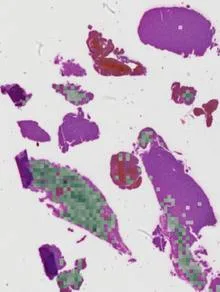
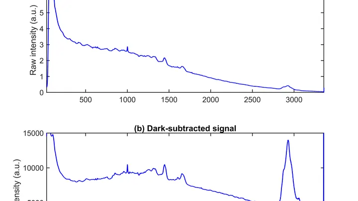
A tissue sample is extracted and then processed in a pathology lab: it is fixed, embedded in paraffin, sliced into thin sections, and stained with dyes that highlight different cellular components. A pathologist examines the slides under a microscope, looking for signs of disease such as malignant cells, inflammation, or infection. In recent years, molecular techniques like geneticsequencing and spectroscopy have been added to enhance diagnostic accuracy, sometimes even analyzing the sample's optical or electrical properties.
In practice
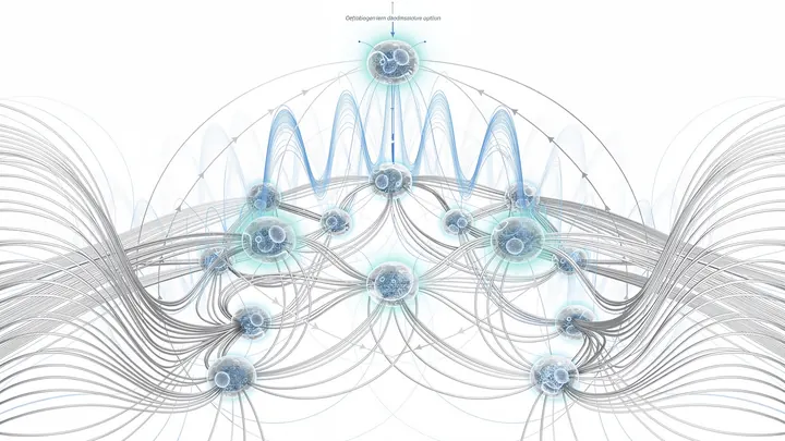
Biopsies are routinely used to diagnosecancer, inflammatory diseases, and infections, and to guide treatment decisions. They are also essential in research, for example in studying how cells communicate via biophotons or how blood biomarkers can reveal disease from a single molecule.
Fun fact
A biopsy sample can be as small as a few hundred cells, yet it can provide enough information to determine the exact geneticmutations driving a tumor.
A stain is found at a crime scene. The expert takes a swab, and a few hours later the instrument outputs a list of proteins that made up the fluid. Previously, a rapid test could be wrong if multiple fluids were present: blood mixed with saliva, and the strip gave a false result.
A sheet of paper tears where tension exceeds strength. Epithelium, the cell sheet lining our organs, behaves similarly, but until recently models could not show how tears appear in it. The classical vertex model describes tissue as a network of polygons tightly packed together and forbids cells from detaching.
Imagine trying to figure out what happened in a room from the sound that lingers after a gunshot. The shot is gone, but the echo still bounces off the walls. That's roughly how a neuropathologist views a brain tissue section where the tumor itself is absent: the biopsy only captured healthy tissue that reacted to the disease.
A doctor looks at a screen and sees two images of the same part of the body. One shows where blood is rich in oxygen, the other shows how fast it rushes through the finest vessels. Previously, these images were made by different devices, and combining them was difficult. Now engineers have built a device that takes both images simultaneously.
A drop of blood from a fingertip lands on the white membrane of a cartridge. Twenty minutes later, the phone shows whether Lyme disease is present or not. No test tube, no centrifuge, no waiting for days.
Usually, for such a test, blood is first spun in a centrifuge to separate the liquid part — the plasma.
A glucose meter that doesn't prick your finger but just looks at your wrist: that's the dream for millions of people with diabetes. It's the promise of algorithms that extract glucose levels from the ordinary pulse signal, the photoplethysmogram (the same one that measures heart rate in smartwatches).
Radiation therapy is a precision strike on a tumor, but every shot must be calibrated to within a millimeter and a fraction of a second. The problem is that existing dosimeters are either bulky, or they distort the beam themselves, or they require pauses for readout.
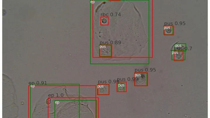
A drop of urine under a microscope isn't a flat puddle but a multi-layered volume. Like a stack of transparent pages: if you focus on one, the neighboring ones blur. The microscope's light only shows one depth layer sharply. To see all the cells, a lab technician had to take a series of snapshots, manually adjusting focus — tedious and slow.
The search for life beyond Earth is like tuning an old radio: we listen to the chemical whisper of distant exoplanets' atmospheres, trying to pick up signs of biological activity.
Diabetes is a constant reminder of needles. To check glycated hemoglobin (HbA1c — a blood protein that reflects average sugar over three months), you need regular blood draws from a vein. It's unpleasant, requires a lab, and takes time. And if you live far from a city, every test becomes a major expedition.
The new iGLU 5.0 device changes the game.
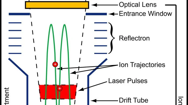
The search for extraterrestrial life is like a detective investigation: from tiny clues, you must distinguish traces of life from random imprints of non-living nature. On Saturn's moon Enceladus, the investigators are lucky — geysers burst from cracks in the icy crust, ejecting water from the subsurface ocean straight into space.
A drop of blood from a vein falls under three beams, and each one drags its own clues into the light. The infrared beam makes molecules vibrate — from that trembling, you learn how much sugar and fat is in the sample. A green laser scatters off proteins and lipids, shifting color — revealing the remodeling of cell membranes.
A drop of ink in a glass of water spreads quickly and evenly. But if the water is squeezed between tightly packed spheres, the ink cloud crawls slowly and erratically: it dodges obstacles and gets stuck in narrow necks. Water molecules in living tissue move in much the same way.
A surgeon removes a breast tumor, but how do you know no cancer cells are left at the edge? Right now, the tissue goes to a lab, and the answer comes back days later. If the margin is not clean, a second surgery is needed. The new method promises to give the answer right in the operating room, while the patient is still on the table.
Your finger is a living sensor: beneath the skin, arteries pulse in time with your heart. Traditional blood pressure cuffs squeeze your arm and give a single number, but this ring uses eight electrodes to pass a weak current through the tissue and map blood flow.
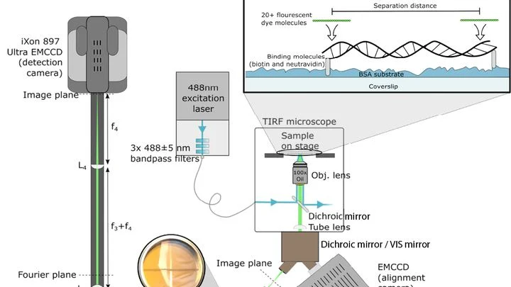
A conventional light microscope hits a fundamental limit: it cannot distinguish two points closer than 200 nanometers — roughly the thickness of a soap bubble divided by a hundred. To see deeper, scientists have made molecules in the sample blink or fade, but these tricks are slow and harmful to living cells.
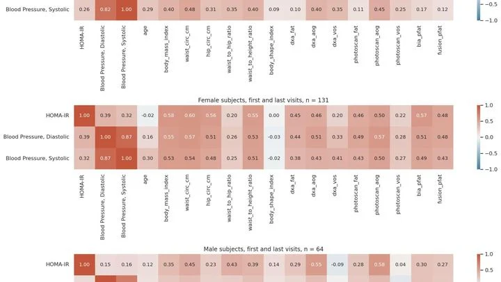
The scale tells you your weight, but it's silent about where those pounds sit — in muscle or belly fat. And it's the fat tucked deep inside, around your organs, that's most often linked to heart disease and diabetes. Previously, you could only see it with an expensive scanner at a clinic. Now, a smartphone is all you need.
In a hospital, blood from a finger prick usually goes to the lab, and the result comes back the next day. The new method promises to cut that wait to a few minutes. Instead of catching light reflected off a sample, it reads the electrical voltage that arises at the boundary between two media, like the faint tingle you feel when you touch a battery with wet fingers.
A scanning tunneling microscope probes a molecule with an ultra-fine tip and maps its relief. But the image is flat, while the molecule is three-dimensional. How do you guess a person's pose from their shadow on a wall?
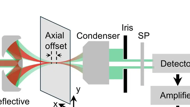
Light we can't see but feel as heat turns out to be the key to cellular life. A new type of microscope uses waves 2000–2500 nanometers long, three times longer than red light, and such waves barely scatter in tissue. Thanks to this, the device peers into a tumor spheroid like a flashlight through frosted glass, illuminating individual molecules.
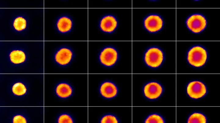
A lightning bolt strikes an anthill. Some ants die instantly, some freeze, and a few days later the survivors start rebuilding the tunnels. That's roughly how a tumor behaves after electroporation — a method where short electric pulses are passed through tissue to poke holes in cancer cell membranes and kill them.
A fat droplet inside a living cell is like a bobber in rough water. It doesn't move in a straight line but in jerks, sometimes pausing, sometimes speeding up. Previously, such movements could only be seen in a single plane, like watching the bobber from above without knowing its depth.
A fingernail-sized piece of ceramic is squeezed in a vise, and its internal structure rearranges like clay under your fingers. Inside, orderly rows of tiny domains line up where they were once randomly oriented. After this 'massage,' the crystal lets light through almost like glass and, at the same time, generates a strong electric charge when bent or pressed.
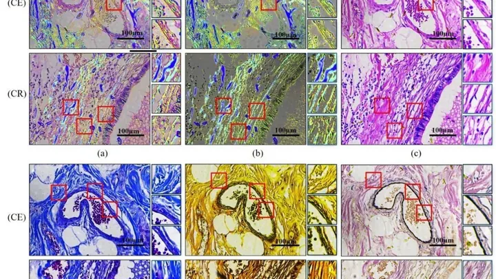
Inside our bodies, between cells, there is a support network — the extracellular matrix. It's like scaffolding made of proteins and carbohydrates that holds cells in place. When the fibers in this network change, it can be a sign of disease — for example, cancer or fibrosis.
Usually, to see these fibers, tissue is cut into thin slices and stained with special dyes.
A tumor doesn't stay put. Individual cells or whole groups break off and set off on a journey through the body. Some crawl solo, like scouts; others stick together, like a flock. Scientists built a computer model to understand how these cells pick a direction.
In the model, cells respond to two signals.
A photographer snaps a runner: to avoid a blurry shot, a short shutter speed is needed. An MRI scanner is like that photographer, but its 'shutter speed' is long — by the time it captures, the muscle has already changed. That's why dynamic MRI (a series of volumetric frames in motion) is a tough challenge: data are scarce, and motion doesn't wait.
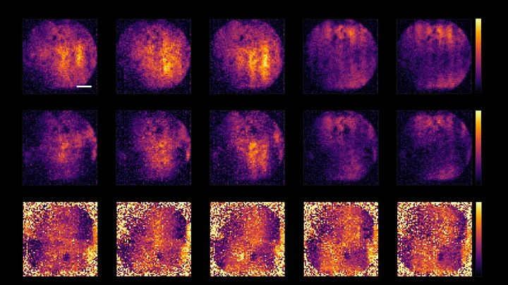
A chemical map of a sample is like a mosaic: each point has its own color, its own chemical composition. To assemble such a mosaic, a spectromicroscope scans the sample point by point, recording a spectrum at each location — a "fingerprint" of how the material absorbs or emits light.
A heart cell in a Petri dish contracts, and at that moment, an electrical wave sweeps across its surface. Previously, to catch that wave, you had to poke the cell with an ultra-fine electrode needle. Now, you just look at it under a microscope.
The method is called electrochromic recording.
In a heart-lung machine, blood flows through narrow tubing and past rapidly spinning pump blades. Sometimes red blood cells can't take it: their membranes rupture, and hemoglobin spills into the plasma. For a patient, this is like pouring sand into a car's fuel system — small damages accumulate and wreck the whole mechanism.
A drop of blood is a bustling city in miniature. Thousands of different molecules scurry through it, each leaving its own trace. But peering directly into this city is hard: the streets are tangled, and there are so many inhabitants that counting them all individually is out of the question.
Scientists have found a way to listen to the whole city at once, without singling out its residents.
Silence in the operating room. The surgeon slowly lowers a needle thinner than a human hair into the patient's brain. At its tip is not a point but a whole palisade of nine hundred sixty microscopic microphones. Each one listens to its own tiny patch of gray matter.
To spot life on other planets, you don't necessarily need to search for oxygen in the air. Living beings paint the surfaces of their worlds: Earth's plants in green, bacteria in purple. The future HWO space telescope will capture these "paints of life" on exoplanets.
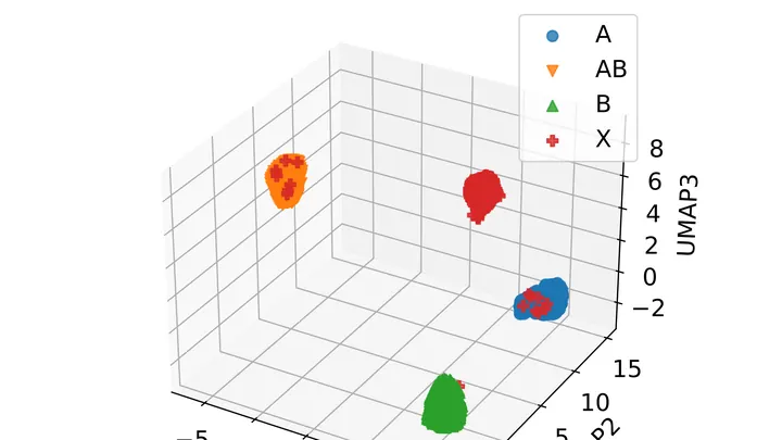
Glioblastoma, a brain tumor, is a tricky foe: in some patients, the drug temozolomide works, while in others it barely helps. The difference often lies in a small stretch of DNA — the MGMT gene. If this region is chemically switched off (methylated), the tumor is worse at repairing its cells, and the drug hits harder.
Living cells continuously exchange light signals — a billion times fainter than daylight. It's like a whisper picked up by ultra-sensitive instruments. A healthy cell whispers rhythmically, a cancerous one — haltingly, but with a long memory of previous flashes.