Steel Under the Microscope: How Manganese Atoms Reveal the Metal's Secrets express
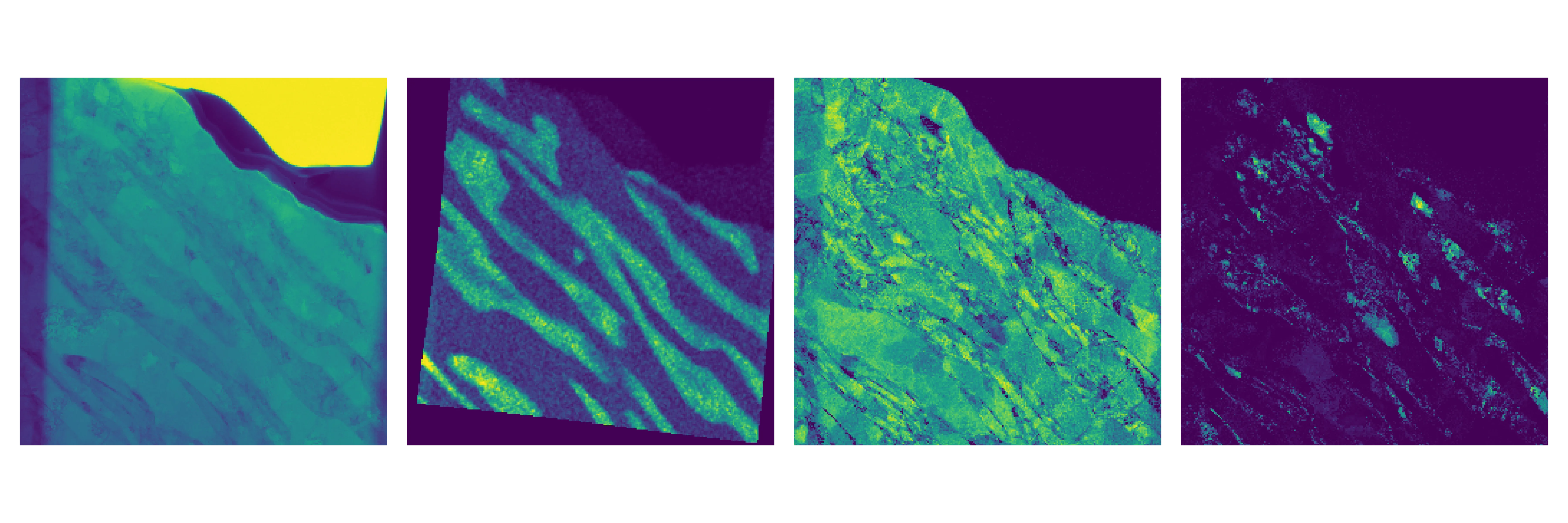
Steel is not just a piece of iron. Inside it, like in a layered pie, different structures, phases, coexist. In ultra-strong steel for cars, there are three such phases: soft ferrite, strong martensite, and ductile austenite. Medium-manganese steel owes its strength precisely to this trio. But how the phases are distributed throughout the volume and how they change under load has long remained a mystery.
Scientists took a sheet of this steel and stretched it like chewing gum. Then they cut ultra-thin slices from both the deformed and untouched areas. Under the electron microscope, they combined two methods: one revealed the crystal lattice, the other mapped the manganese distribution. This allowed them to see, for the first time, which phase is where and how much manganese it contains, with a precision of 10 nanometers — a thousand times thinner than a human hair.
The results were surprising: after stretching, the metal's grains became finer, and their orientation changed. Ferrite turned out to be less stressed than martensite. Austenite partially transformed into martensite: this is what gives the steel its ultra-high strength. Now engineers can more precisely tailor the steel's composition to make cars lighter and safer.
Original abstract
as written by the authors arXiv:2601.22407
The mechanical response of third-generation advanced high-strength steels is governed by phase transformations at the nanoscale, yet the coupled evolution of chemistry and crystallography remains poorly resolved. Here we apply a correlative scanning transmission electron microscopy approach that enables simultaneous mapping of lattice structure, crystallographic orientation, and phase distribution at 10 nanometre resolution in a medium-manganese TRIP steel. We combine nano-beam electron diffraction and energy-dispersive X-ray spectroscopy maps to characterize an industrial medium-manganese steel containing 7.15 weight percent Mn. Tensile testing of a rolled steel sample was performed, and lamellae were extracted from deformed and undeformed regions. Manganese-resolved energy-dispersive X-ray spectroscopy provides a chemical fingerprint that, when combined with nano-beam electron diffraction based phase segmentation, enables robust ferrite-martensite separation and phase-resolved lattice-parameter refinement. The phase fractions of ferrite, austenite, and martensite are quantified together with their corresponding lattice parameters, accompanied by measurable shifts in grain-size distributions and crystallographic texture in the deformed regions. Kernel average misorientation maps reveal systematically lower local misorientation in ferrite than in martensite. This multimodal workflow provides a transferable framework for quantitative, phase-resolved analysis of complex multiphase alloys at the nanoscale.
In one cubic centimeter of such steel, there are so many grain boundaries that their total area is comparable to that of a football field.