How to Assemble an Aorta from Six Scans express
The body's main artery, the aorta, is not just a tube. With each heartbeat, it stretches and contracts like a rubber hose under water pressure. Seeing this motion in volume is hard: standard MRI gives only flat slices, and full 3D imaging over time requires expensive equipment and long periods lying in the scanner.
A group of researchers found a way out. They built a "library" of hundreds of typical aortic shapes, like a tailor's pattern catalog. Then they took just six flat scans from a specific patient and used an algorithm to pick the shape from the library that best matches those slices. The algorithm is like a sculptor who reconstructs an entire statue from a few contours.
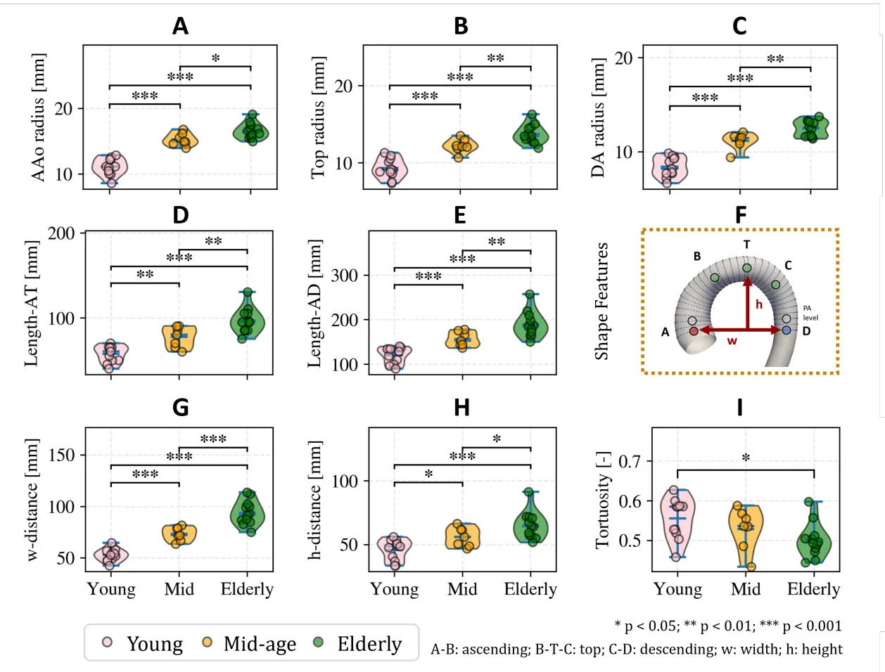
Tests on 30 volunteers and patients showed that age changes the aorta's elasticity. In young people, the wall stretches 11% with each beat; in older people, only 3%. That's the difference between a fresh rubber band and an old, cracked one.
This approach makes a 3D view of the aorta accessible in regular clinics: no new equipment needed, just smart processing of familiar scans. And that means doctors can spot dangerous changes in the main artery earlier.
Original abstract
as written by the authors arXiv:2602.11873
Background and Objective: To assess the feasibility and accuracy of reconstructing time-resolved, three-dimensional, subject-specific aortic geometries from a limited number of standard cine 2D magnetic resonance imaging (MRI) acquisitions. This is achieved by coupling a statistical shape model with a differentiable volumetric mesh optimization algorithm. Methods: Cine 2D MRI slices were manually segmented and used to reconstruct subject-specific aortic geometries via a differentiable mesh optimization algorithm, constrained by a statistical shape model. Optimal slice positioning was first evaluated on synthetic data, followed by in-vivo acquisition in 30 subjects (19 volunteers and 11 aortic stenosis patients). Time-resolved aortic geometries were reconstructed, from which geometric descriptors and radial strain were derived. In a subset of 10 subjects, 4D flow MRI data was acquired to provide volumetric reference for peak-systolic shape comparison. Results: Accurate reconstruction was achieved using as few as six cine 2D MRI slices. Agreement with 4D flow MRI reference data yielded a Dice score of (89.9 +/- 1.6) %, Intersection over Union of (81.7 +/- 2.7) %, Hausdorff distance of (7.3 +/- 3.3) mm, and Chamfer distance of (3.7 +/- 0.6) mm. The mean absolute radius error along the aortic arch was (0.8 +/- 0.6) mm. Secondary analysis demonstrated significant differences in geometric features and radial strain across age groups, with strain decreasing progressively with age at values of (11.00 +/- 3.11) x 10-2 vs. (3.74 +/- 1.25) x 10-2 vs. (2.89 +/- 0.87) x 10-2 for the young, mid-age, and elderly groups, respectively. Conclusion: The proposed framework enables reconstruction of time-resolved, subject-specific aortic geometries from a limited number of standard cine 2D MRI acquisitions, providing a practical basis for downstream computational analysis.
The aorta is the largest blood vessel in the human body: in an adult, its diameter at the root reaches 3–4 cm, about the size of a garden hose.
In the series "The Expanse," ship doctors use portable scanners that instantly build 3D models of internal organs: this method takes a step toward such compact diagnostics.