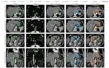
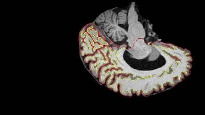
Medical image segmentation automatically divides a scan into anatomical or pathological structures. Modern methods, especially deep learning architectures like U-Net, process images at multiple scales, capturing both fine details and overall context. The output is a probability map for each pixel—the chance of belonging to one class or another (tumor, liver). Training requires large annotated datasets, which are often augmented artificially (rotations, flips).
History
Threshold-based segmentation emerged in the 1970s. In 1988, Kass et al. proposed active contour models that optimize energy functions to find boundaries. With deep learning, the U-Net architecture (Ronneberger et al., 2015) revolutionized the field, enabling end-to-end training on limited data. Subsequent improvements include attention mechanisms and transformer-based models. Leading conferences: MICCAI, MIDL.
How it works
A CNN is trained on images with pixel-wise annotations. U-Net compresses the image into a bottleneck through convolutional and pooling layers, then expands it back with transposed convolutions. Skip connections concatenate high-resolution encoder features with the decoder, preserving fine details. The final layer uses softmax (for multiple classes) or sigmoid (for binary cases). Dice loss (1 - 2|X∩Y|/(|X|+|Y|)) handles class imbalance. Post-processing (connected components, CRF) refines boundaries.
In practice
Outlining tumor boundaries on MRI for radiation therapy planning or measuring organ volumes on CT.
Fun fact
The U-Net architecture, shaped like the letter 'U', won the ISBI cell tracking challenge in 2015 and now underlies most medical segmentation tools.
A mapmaker drawing a route through a city doesn't sketch every brick in every wall. Streets, intersections, and distances are enough. The same goes for calculating radiation dose in breast tomography: you don't need every cell, just a faithful overall picture.
A radiation oncologist looks at an abdominal scan and mentally outlines the area to be irradiated. That area is not visible as a separate organ: its boundaries are set by protocol, not by tissue. So the doctor needs a map of the surroundings to avoid hitting healthy structures.
The nnU-Net model starts with the same task.
A radiation oncologist looks at an abdominal scan and mentally outlines the area to be irradiated. That area is not visible as a separate organ: its boundaries are set by protocol, not by tissue. So the doctor needs a map of the surroundings to avoid hitting healthy structures.
The nnU-Net model starts with the same task.
A thin section of eucalyptus lies under the microscope objective, and white light, passing through two polarizing filters, turns the wood into a mosaic of purple, yellow, and blue patches. These colors are not random: like letters on a map, they show the direction of the invisible cellulose threads inside the cell walls.
A radiologist looks at an abdominal scan and sees not just organs but their spatial relationships: which kidney is larger, where the tumor lies relative to a vessel. It's like reading a map, where it's not enough to know where a city usually stands; you also need to measure the distance between two points on the ground.
A radiologist looks at an abdominal scan and sees not just organs but their spatial relationships: which kidney is larger, where the tumor lies relative to a vessel. It's like reading a map, where it's not enough to know where a city usually stands; you also need to measure the distance between two points on the ground.
A doctor looks at a screen and sees two images of the same part of the body. One shows where blood is rich in oxygen, the other shows how fast it rushes through the finest vessels. Previously, these images were made by different devices, and combining them was difficult. Now engineers have built a device that takes both images simultaneously.
You're looking for fireflies in a dark forest. Instead of photographing the whole forest every second and then poring over the images, you carry a small device that spots the flashes on its own and cuts out only a circle around each one. The rest, the blackness, isn't saved. That's exactly how the new sensor for scientific cameras works.
A stenographer is recording a meeting, but halfway through the conversation she runs out of paper. She misses a few lines, then picks up where she left off. Later, she needs to reconstruct the full transcript without rewriting the entire conversation from scratch. That's exactly how the new method URA-SIM works for structured illumination microscopy (SIM).
A shipping container is a steel safe the size of a room, packed with goods. X-rays struggle to see through it: thick walls and dense cargo block the rays. Enter muons — particles born high in the sky that constantly rain down on Earth. They can pass through even lead, but they veer slightly off course when they encounter something heavy.
Fatty liver disease often goes unnoticed, but over time it can lead to serious problems. To avoid taking a tissue sample, doctors use ultrasound: sound waves pass through the tissue and attenuate differently depending on fat content. This parameter, the attenuation coefficient, shows how "fat" the liver is.
But the method has a flaw.
A tumor in a scan is like a tiny bright dot on a vast dark canvas. That dot is what the doctor needs, not a perfectly smooth background. But when a neural network learns to turn a regular CT scan into something resembling a PET scan, it often tries to make everything look nice, and ends up smearing that dot.
Two scans of the same brain taken at different times are like photographs of the same house before and after renovation. To see what changed, you overlay them and look at the differences. But an MRI scanner is not a perfect camera: it adds its own distortions, as if the photo were taken through slightly warped glass.
A doctor spends years learning to recognize diseases from brain scans. First, they excel at spotting signs of aging, then they master tumors, and suddenly they start confusing what they knew before. Many neural networks behave much the same way: new knowledge overwrites the old.
A flashlight shines through frosted glass: the rays bend, lose brightness, and take different paths. That's roughly how high-intensity ultrasound passes through the skull to the brain. For years, calculating this path required bulky computer models that 'chew' on data for hours.
An X-ray machine takes a scan, and the doctor sees only numbers — bone density, fat percentage. But the image holds far more. A team of researchers created a model called LeDXA that reads these scans like a map of hidden bodily signals.
The model was trained on 11,540 scans from the Human Phenotype Project with no labels at all.
A huge orchestra without a conductor. Each musician is a gene, and the melody is the cell's complex work. Previously, looking at a tissue photo, a computer tried to guess each instrument's part individually. It didn't know that violins usually play with violas, and trumpets with trombones. The program just output a list of notes, without sensing the harmony.
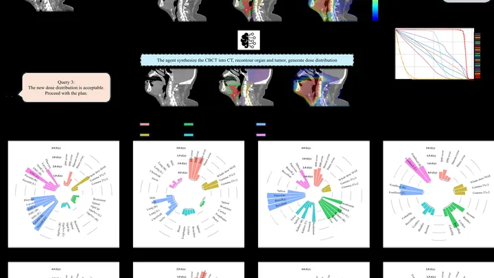
The proton beam is like a sniper's bullet: it delivers most of its energy at a precisely set point, barely touching tissues on the way in. But over 4–6 weeks of head and neck tumor treatment, the target shifts: the patient loses weight, the tumor shrinks, posture changes. The sniper aimed at one spot, but the target has already moved.
The brain is an orchestra where each group of instruments must come in on time. In Alzheimer's disease, the musicians lose their notes: connections between brain regions get tangled, and the harmonious performance turns into cacophony. Scientists have long seen these disruptions on scans, but they didn't know if the situation could be fixed by touching just one string.
To capture every detail of a statue's shape, you could break it into millions of tiny cubes and describe each one — slow, heavy, and your computer's memory would groan at the seams. That's how 3D medical scans are usually handled: a CT or MRI is a huge array of such cubes (voxels), and to create or improve an image, the machine has to plow through all of them.
A single-pixel camera works like an artist creating a picture through a stencil with many holes. In each frame, only a portion of the light, modulated by a mask, a spatial light modulator, reaches the single sensor, and then the computer reconstructs the full image by assembling this mosaic with computational algorithms.
Each morning, a navigator plots a route, bypassing traffic jams and roadblocks. Similarly, before a radiation therapy session, a new AI agent recalculates the irradiation path.
Ordinary AI looks at medical images like puzzles made of unfamiliar pieces: it considers any two unmatched pictures completely different. It's like trying to tell twins apart without knowing they're related. In medicine, however, patients with the same condition look nearly identical on MRI, and the algorithm mistakes 'relatives'.
Genes are like houses in a city: in some, life bustles; in others, silence reigns. This 'urban' genetic landscape was analyzed in a thousand women with breast cancer.
Imagine a kinked garden hose: water meets resistance, and pressure drops. A similar process occurs in arteries when they lose their straight geometry. Vascular tortuosity is not just a cosmetic defect but a factor that disrupts hemodynamics and poses a threat of ischemic stroke.
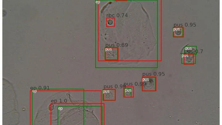
A drop of urine under a microscope isn't a flat puddle but a multi-layered volume. Like a stack of transparent pages: if you focus on one, the neighboring ones blur. The microscope's light only shows one depth layer sharply. To see all the cells, a lab technician had to take a series of snapshots, manually adjusting focus — tedious and slow.
The data from a single CT scan is comparable to several feature-length films. TCAM-Diff bypasses this bottleneck: the program compresses the 3D scene into three shadow projections — like a sculptor reconstructing a solid statue from silhouettes on three walls.
Ultrasound waves bounce off organs to draw their outlines, but on an ovarian scan it's hard to tell a harmless fluid-filled cyst from a solid cancerous mass. Even specialists draw the boundaries differently, and this directly affects treatment choices.
The OvAi Focus program tackles this by learning from thousands of examples.
Usually a CT scanner takes many X-ray images from different angles and assembles a 3D picture. But sometimes doctors only scan a small area (say, one lung) and the data cuts off. It's like a chef given a single spoonful of soup and asked to name the exact recipe for the entire pot.
Medical scans are maps of tissue density. The brightness of each pixel reveals what’s there, much like photometry measures light intensity. But conventional algorithms analyze pixels in isolation, producing jagged organ boundaries.
A new approach compels the program to prioritize smoothness.
In a noisy room, a person picks out a desired voice thanks to two ears capturing the difference in sound arrival. A computer with several microphones does the same: it gets spatial cues — where each voice is coming from.
Previously, these cues were used to focus on one direction, like a flashlight beam.
Imagine photographing a statue in dim light. You face a dilemma: take one bright shot or a series of dim frames from different angles. In the first case, quality is limited by photon noise, the graininess that arises from a lack of photons, while in the second, details overlap, making it harder to reconstruct the 3D structure.
Tomographs don't produce ready-made pictures. They collect numerous 'shadows' — projections of innards from different angles or using various signals — and mathematically reconstruct a 3D picture from them.
In brachytherapy, thin catheters are inserted into tumors. On CT scans, they look like tangled glowing threads. Previously, doctors would manually outline each one, spending up to an hour.
The new algorithm learns from synthetic scans — drawing metal strips in an empty volume, like threads without a body.
Prostate MRI scans often look like photos taken through a dirty window: noise, blurred edges, curvature from field imperfections. Scientists checked what happens if you 'wash' that window: remove the excess, correct the distortions, and accurately align different types of scans.
The eye is inflamed, the cornea clouding. The doctor needs to quickly determine the culprit — bacteria or fungus. The treatment depends on it. A mistake can cost sight. But the classic lab answer takes days, and in remote areas it may never arrive.
Researchers from the US and India propose a different approach: three images from a slit lamp — a device found in nearly every eye clinic.
A radiologist stares at gray-white smudges on the screen — dozens of body slices where every suspicious spot must be found. Manual segmentation of one patient takes half an hour, and a large clinic sees hundreds of such patients. Fatigue and varying experience inevitably introduce variability: two specialists will outline the same lesion slightly differently.
Look at an equilateral triangle — the probability simplex for three assets. Each interior point is a portfolio: non-negative shares summing to one. The triangle's center, the barycenter, corresponds to an equal-weight allocation (1/3, 1/3, 1/3). It seems that by moving toward this point, you lower risk and avoid dangerous edges.
The heart sometimes loses its rhythm, like an orchestra where the violins suddenly start playing each on their own. Cardiologists in such cases look for the "out-of-tune" areas by reading the heart's electrical signals and precisely ablating them with a thin probe — a catheter.
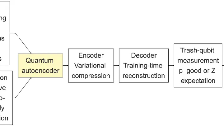
A cook spends years honing the skill of reducing a recipe to three or four essentials and recreating the dish from them. If the ingredients are familiar, the expected meal lands on the plate. A foreign ingredient disrupts compression, and you get something unrecognizable. The Quantum Compressor does the same with medical scans.