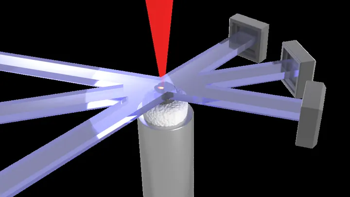
X-ray imaging optimization is the field of improving the quality of X-ray images while minimizing the radiation dose delivered to the subject. It is a constant balancing act: higher radiation doses generally produce clearer images, but they increase the risk of cellular damage, especially in medical diagnostics. The field encompasses everything from hardware design (better detectors, more efficient X-ray sources) to software algorithms (noise reduction, image reconstruction) and even novel imaging geometries that capture more information per exposure.
History
The field began with Wilhelm Röntgen's discovery of X-rays in 1895, but systematic optimization efforts started in the mid-20th century as the risks of radiation exposure became understood. The introduction of computed tomography (CT) in the 1970s by Godfrey Hounsfield and Allan Cormack brought a new dimension to optimization, as CT inherently requires many exposures.
How it works
Traditional X-ray imaging uses a camera or film to capture the shadow cast by an object illuminated with X-rays. Optimization techniques work at multiple levels. Hardware improvements include more sensitive detectors that capture a higher fraction of incoming photons, and better X-ray optics that focus or collimate the beam to reduce wasted radiation. Software methods use statistical models to separate true signal from noise, allowing lower-dose images to be 'cleaned up' after capture. Some advanced approaches, like ghost imaging or computational imaging, reconstruct images from sparse or indirect measurements, extracting maximum information from minimal radiation.
In practice
Used in medical diagnostics (CT scans, mammography, dental imaging) to reduce patient dose while maintaining diagnostic accuracy, and in materials science for non-destructive testing of components where repeated imaging is needed.
Fun fact
Some modern X-ray imaging techniques can reconstruct a 3D image of a rotating object from a single exposure by using coded apertures, effectively encoding depth information in the diffraction pattern.
A translator at the negotiating table doesn't listen to every word; they grasp the meaning of whole phrases. Electron microscopy poses a similar challenge: you need to figure out the crystal's thickness and its tilt in the beam, and all you have is a diffraction pattern, a mosaic of thousands of bright and dark spots.
A globe sits on a table, and a ruler lies beside it: one is curved, the other straight. X-ray telescopes have lived with this mismatch for years. Their mirrors gather light onto a curved surface, like the globe, while the detectors that catch that light have been flat, like the ruler.
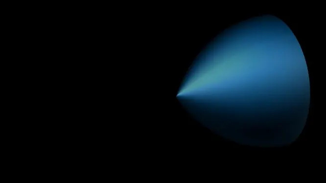
A gas target is like a funnel: the steeper its walls, the stronger the flow. But in this funnel, it is not water that flows, but ultraviolet light, born when a powerful laser pulse passes through a cloud of argon. The efficiency of this conversion is usually minuscule, about 0.001%: out of a thousand laser photons, only one turns into useful ultraviolet.
A smartphone camera captures light with tiny sensors, but by themselves, they can't distinguish colors. So color filters are placed in front of them: red, green, blue, like stained glass in a window. Each sensor sees only its own color, and then software assembles the full picture from these pieces.
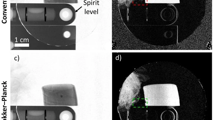
A low-dose X-ray image looks like a view through a fogged-up window: outlines are there, but details drown in a hazy veil. Doctors need such images during radiation therapy — to aim the beam precisely at the tumor and spare healthy tissue. But saving on radiation comes at the cost of noise, streaks, and artifacts that obscure what matters.
The Sun is a giant cauldron where light struggles through a thick layer of scorching matter. On its way out, photons (light particles) constantly bump into atoms and lose energy. How strongly matter holds back light is called "opacity," and it determines how quickly heat escapes.
Iron is one of the main culprits for opacity in the Sun's interior.
A camera flash that fires in a thousandth of a second doesn't heat the subject — unlike a regular flash that lasts a full second. A similar trick has been found in radiotherapy: when a radiation dose is delivered in milliseconds instead of minutes, healthy tissues suffer less. This is called the FLASH effect.
Imagine photographing a statue in dim light. You face a dilemma: take one bright shot or a series of dim frames from different angles. In the first case, quality is limited by photon noise, the graininess that arises from a lack of photons, while in the second, details overlap, making it harder to reconstruct the 3D structure.
Dozens of copper coils in an MRI scanner pick up the faint response from tissues, like microphones trying to catch a whisper in a noisy hall. Each microphone drags a thick bundle of wires behind it. The wires press on each other, create electrical interference, heat up, and add risk for the patient.
A powerful laser strikes a diamond like a sledgehammer, with matter itself serving as the anvil. The impact lasts only an instant but generates pressures a million times greater than atmospheric. Physicists have long dreamed of peering into that moment to see how atoms rearrange under the onslaught.
The X-ray beam here is like a camera flash.
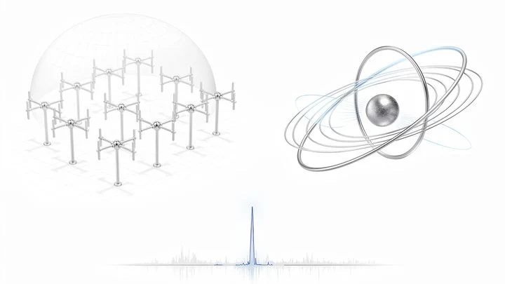
Listening to the cosmos requires patience and a finely tuned ear. Astronomers chose the most sensitive low-frequency instrument for this: the OVRO–LWA antenna array — 352 dipole antennas scattered across two and a half kilometers of California desert.
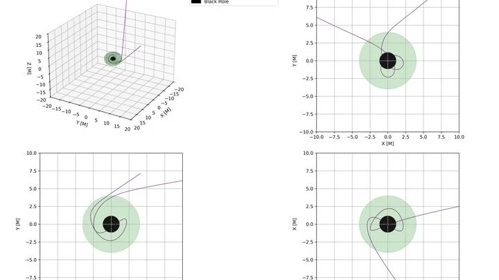
Light passing by a black hole falls into a gravitational trap: its trajectory curves and the particle bounces along a nearly closed orbit, like a pinball.
A flashlight beam aimed through frosted glass turns into a blurry spot with every extra millimeter of thickness. Scintillators, materials that convert X-rays into visible light, behave much the same way. They underpin medical imaging, baggage screening, and detectors at particle accelerators.
Any light measurement runs into a fundamental limit: photons hit the camera sensor unevenly. This natural ripple (shot noise) blurs fine details, like trying to pick out a quiet instrument in an orchestra through the noise of the hall.
An X-ray machine with gratings takes not one but three pictures at once: a regular image, a map of beam deflections invisible to the eye, and an image of tiny structures that scatter light. Together, they help spot, for example, emphysema or pulmonary fibrosis. But there's a catch: to separate these three layers, you need a clever mathematical trick.
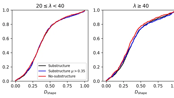
Giant galaxy clusters are cities of thousands of star islands, held together by gravity. Inside them, hot gas churns and glows in X-rays. If a cluster has recently collided with another, it develops clumps of galaxies and distortions in the gas, like ripples on water after a stone is thrown.
Light passing through a turbid medium like milk or living tissue scatters in all directions. Reconstructing where light was absorbed more strongly is like guessing the shape of a fallen object from a puddle on the asphalt. Optical tomography solves this problem, but usually requires large computational resources.
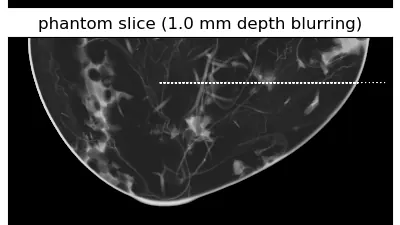
The X-ray machine moves around the breast in an arc, taking several images from different angles. From these flat frames, the computer assembles a 3D picture, like putting together a mosaic from pieces. But if the mosaic pieces are crumpled or smudged, the picture turns out blurry. In tomography, the role of "crumples" is played by noise and imperfections in the imaging.
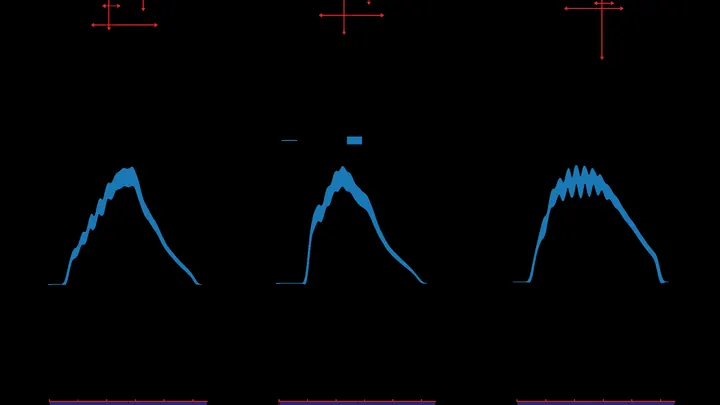
A tomograph is like a photographer who walks around an object and takes pictures from different sides. Usually, it snaps at regular intervals and uses the same amount of light for each shot. But if you need to shoot quickly and not waste extra film, you have to choose: which angles matter more and where to direct more light.
The neutron star 4U 1735-44 is gobbling up matter from a companion star. The matter spirals into a flat disk, like water going down a bathtub drain. But this disk shines in X-rays, and astronomers have for the first time measured how "combed" those rays are: that is, how polarized.
A thin film for a solar cell or display is like a dense crowd in a square: each person faces a different direction, and whether light passes through the crowd or gets stuck depends on which way everyone is looking. In organic electronics, molecules play the role of passersby. Until now, instruments could only measure the crowd's overall direction — the average head tilt and its spread.
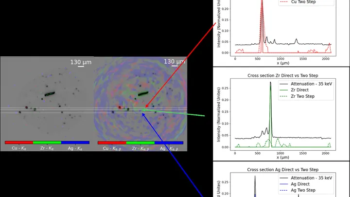
Conventional X-ray fluorescence analysis works like a meticulous scribe: to learn the composition of a picture, it walks over every square centimeter and logs whether lead or copper is present. For a flat sheet, that's tolerable, but for a three-dimensional object, it's torture.
Quantum annealing works like a smart way to find a path through a maze: instead of checking every dead end, it uses quantum effects to find the exit faster. But this maze has a quirk: the numbers that describe the walls and corridors must be roughly the same size.
In the operating room, the surgeon illuminates the tumor with a special agent that makes it glow. But how do you know whether it glows brightly because there are many cancer cells or simply because it lies close to the surface?
A weak magnet in an MRI machine is like a dim bulb in a dark room: you get a picture, but details drown in grain. These scanners operate at fields below 0.1 tesla (a typical hospital machine runs at 3 tesla, 30 times stronger), yet they're lightweight, inexpensive, and don't require a special room. The catch: noise eats away at quality.
An X-ray image is like a photo taken in the dark: the less light, the harder it is to see details. Doctors wanted to know how much you can dim the "light" in computed tomography (CT) and still not miss a disease. To do this, they took 50 pairs of chest images: one with a full radiation dose, the other with a low dose.
A typical accelerator is like an assembly line where electrons are the workpieces. To get the desired X-ray pulse, the workpiece is fine-tuned after it's already on the line: squeezed, filtered, corrected. In the new work, engineers decided to take a different approach — shaping the workpiece before it even hits the conveyor.
At LCLS-II, electrons are knocked out of a metal by a short laser pulse.
A laser melts metal powder layer by layer, and inside the boiling pool, vapor bubbles appear and collapse within milliseconds. Seeing this kitchen in action was previously impossible: the X-ray tomograph that takes 3D images is too slow. The sample has to rotate, and by the time it does, the melt has already changed a hundred times.
A beam of carbon ions strikes the tumor precisely, like a surgeon who can only see the skin. Inside the body, the beam stops, and exactly where used to be a mystery. Now, helium is mixed into the same beam: it passes through the body like an X-ray, and its trail shows where the carbon went.
The laser beam in a three-photon microscope works like a flashlight shining through murky water. The deeper you need to illuminate, the more the water scatters light and the dimmer the picture gets.
An X-ray image usually brings to mind a bulky machine in a hospital. But such a picture can be taken by a device the size of a palm, assembled from hobbyist electronics. The authors took a Raspberry Pi mini-computer, a 12.3-megapixel camera, and a screen that glows under X-rays.
An X-ray image of a battery looks like a photo taken through frosted glass: fine details blur into a gray smudge. The culprit is the detector itself, which cannot distinguish features smaller than its pixel size. Engineers proposed getting around this barrier by illuminating the sample not with a uniform beam but with a striped pattern.
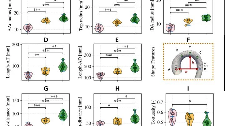
The body's main artery, the aorta, is not just a tube. With each heartbeat, it stretches and contracts like a rubber hose under water pressure. Seeing this motion in volume is hard: standard MRI gives only flat slices, and full 3D imaging over time requires expensive equipment and long periods lying in the scanner.
A group of researchers found a way out.
Our body's tissue is like a city: different neighborhoods are home to different cells, each with its own job. To understand how this city is organized, biologists take images showing which genes are active at each spot. But sharp images are expensive, and cheap ones come out blurry, like a photo of a city from high above, where individual buildings are hard to make out.
In winter, puddles freeze, and ice cracks the asphalt. Inside a living cell, freezing does something similar: ice crystals tear its membrane. To prevent this, the cell is soaked in a special solution, like antifreeze for a car. But precision is key: too little solution and ice still forms; too much and the solution itself poisons the cell.
You can't catch X-rays from stars with an ordinary mirror: they pass through glass like light through a window pane. To reflect them, you need a mirror almost parallel to the beam: then the X-rays skim along the surface like a flat stone skipping on water, only slightly changing direction.
A photo taken through frosted glass always comes out blurry. As light passes through a sample, it scatters, and the camera sees only a smudge instead of a sharp image. Scientists have long known how to recover an image from such a smudge, but old methods are finicky: they either get stuck on a wrong solution or require too many exposures, bombarding the fragile sample.
A heated floor in a room always vibrates slightly — molecules in it are moving. The same happens to mirrors in gravitational wave detectors: heat makes their surfaces tremble finely, which hinders picking up faint signals from space.
To calm the jitter, thin layers of different materials are applied to the mirror, like a layered cake.
Every time an MRI scanner measures water movement in tissues, it sends a magnetic pulse in a specific direction. One pulse gives one data point. To understand how water moves in all directions at once, you need many such pulses with different rotations. Previously, rotations were chosen almost at random, just to cover all directions evenly.
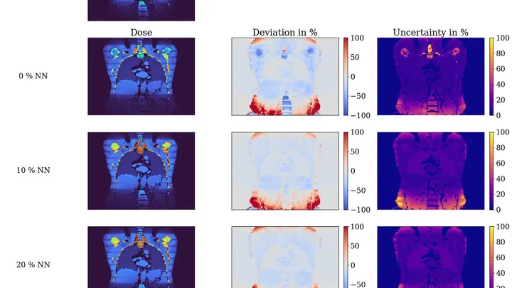
Computed tomography is like an X-ray that takes hundreds of images from different angles and stitches them into a 3D picture of the body. But each image delivers a small dose of radiation, and doctors need to know how much each organ receives. Currently, this is often estimated roughly using average tables rather than for the specific patient.