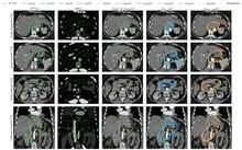
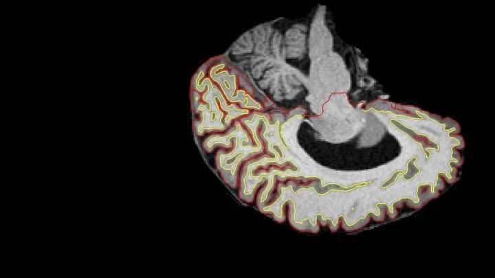
Tumor segmentation is a specific task in medical image analysis where AI algorithms outline the exact boundaries of tumors in medical images. This is crucial for treatment planning, such as radiation therapy, where doctors need to target the tumor precisely while sparing healthy tissue. Segmentation can be performed on various imaging modalities, including MRI, CT, and ultrasound, and across different body parts like the brain, prostate, or breast.
History
Early segmentation methods relied on manual tracing or classical image processing techniques like thresholding and region growing. The introduction of fully convolutional networks around 2015 revolutionized the field, enabling automatic and more accurate segmentation.
How it works
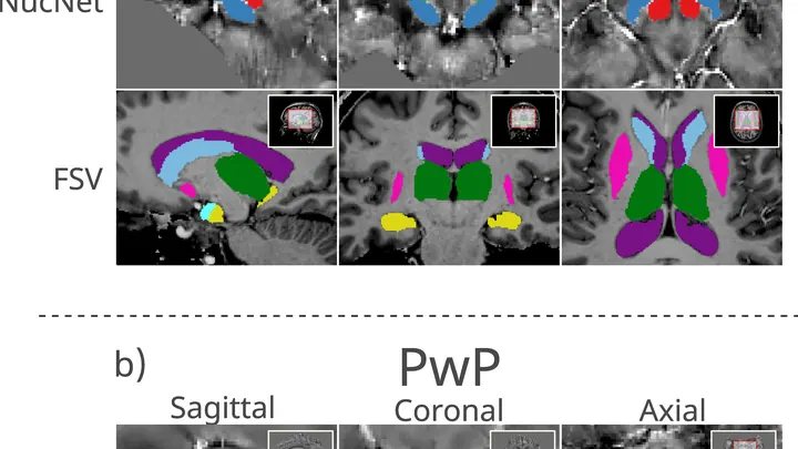
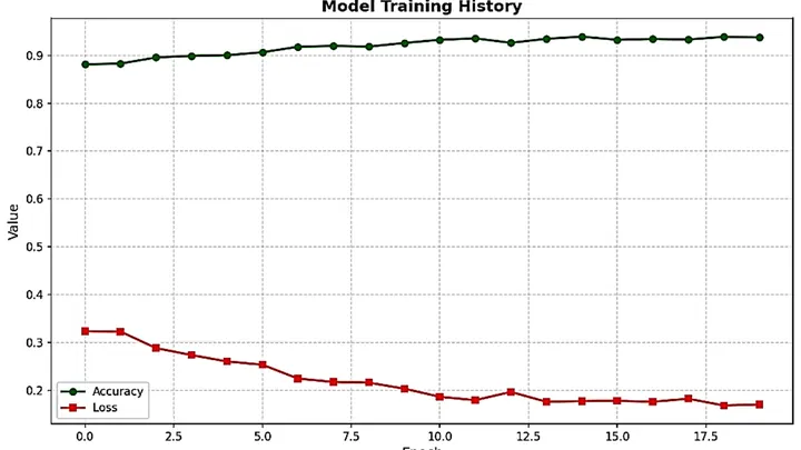
Segmentation models, often based on U-Net architectures, take an image as input and produce a pixel-wise mask that labels each pixel as tumor or non-tumor. The network is trained on images where the tumor boundaries have been manually annotated by experts. Through training, the model learns to recognize the visual characteristics of tumor boundaries, such as irregular shapes, intensity contrasts, and texture differences. Advanced models can also incorporate spatial context from 3D volumes, improving accuracy.
In practice
Tumor segmentation is used in radiotherapy planning, surgical navigation, and monitoring tumor growth over time, as seen in studies on prostate cancer and glioblastoma.
Fun fact
Some segmentation algorithms can work with 'lensless' imaging, reconstructing tumor shapes from shadows in a single image, as demonstrated for breast cancer detection.
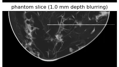
A mapmaker drawing a route through a city doesn't sketch every brick in every wall. Streets, intersections, and distances are enough. The same goes for calculating radiation dose in breast tomography: you don't need every cell, just a faithful overall picture.
A radiation oncologist looks at an abdominal scan and mentally outlines the area to be irradiated. That area is not visible as a separate organ: its boundaries are set by protocol, not by tissue. So the doctor needs a map of the surroundings to avoid hitting healthy structures.
The nnU-Net model starts with the same task.
A radiologist looks at an abdominal scan and sees not just organs but their spatial relationships: which kidney is larger, where the tumor lies relative to a vessel. It's like reading a map, where it's not enough to know where a city usually stands; you also need to measure the distance between two points on the ground.
At the end of every chromosome sits a cap made of repeating DNA letters: the telomere. For a long time it was measured as an average length across all chromosomes, as if judging a library by the average thickness of its books. But in humans, three ends, 17p, 20q, and 12p, are consistently shorter than the rest, and they are the ones more often linked to genome damage.
A stenographer is recording a meeting, but halfway through the conversation she runs out of paper. She misses a few lines, then picks up where she left off. Later, she needs to reconstruct the full transcript without rewriting the entire conversation from scratch. That's exactly how the new method URA-SIM works for structured illumination microscopy (SIM).
A shipping container is a steel safe the size of a room, packed with goods. X-rays struggle to see through it: thick walls and dense cargo block the rays. Enter muons — particles born high in the sky that constantly rain down on Earth. They can pass through even lead, but they veer slightly off course when they encounter something heavy.
High-frequency sound focused on a single point acts like an invisible lens gathering sunlight. Inside a tumor, such a lens can raise the temperature to about 72°C in a second, like hot water from a tap, but inside living tissue. Meanwhile, surrounding healthy cells remain almost untouched.
Two scans of the same brain taken at different times are like photographs of the same house before and after renovation. To see what changed, you overlay them and look at the differences. But an MRI scanner is not a perfect camera: it adds its own distortions, as if the photo were taken through slightly warped glass.
A doctor spends years learning to recognize diseases from brain scans. First, they excel at spotting signs of aging, then they master tumors, and suddenly they start confusing what they knew before. Many neural networks behave much the same way: new knowledge overwrites the old.
A flashlight shines through frosted glass: the rays bend, lose brightness, and take different paths. That's roughly how high-intensity ultrasound passes through the skull to the brain. For years, calculating this path required bulky computer models that 'chew' on data for hours.
A bell hidden in a thick box can be made to ring without opening the lid. Just send a sound wave through the walls, and the clapper will tremble. That's roughly how transcranial ultrasound works: sound passes through the skull bones and makes neurons deep in the brain 'ring'—generating electrical impulses.
An ordinary flashlight emits light whose waves oscillate in all directions at once. But pass that light through a special filter, and you get a beam that oscillates strictly in one plane: that's polarized light. When such a beam travels through biological tissue, the tissue's internal structure slightly alters the orientation of the oscillations.
A pebble on a streambed lies still as sunlight sparkles dance on the water's surface. In astronomy, the contrast is similar: a real exoplanet is the stone, while myriad speckles (patches of scattered starlight) are the ripples trembling in instruments.
Genes are like houses in a city: in some, life bustles; in others, silence reigns. This 'urban' genetic landscape was analyzed in a thousand women with breast cancer.
Ultrasound waves bounce off organs to draw their outlines, but on an ovarian scan it's hard to tell a harmless fluid-filled cyst from a solid cancerous mass. Even specialists draw the boundaries differently, and this directly affects treatment choices.
The OvAi Focus program tackles this by learning from thousands of examples.
Imagine a kinked garden hose: water meets resistance, and pressure drops. A similar process occurs in arteries when they lose their straight geometry. Vascular tortuosity is not just a cosmetic defect but a factor that disrupts hemodynamics and poses a threat of ischemic stroke.
Medical scans are maps of tissue density. The brightness of each pixel reveals what’s there, much like photometry measures light intensity. But conventional algorithms analyze pixels in isolation, producing jagged organ boundaries.
A new approach compels the program to prioritize smoothness.
Tomographs don't produce ready-made pictures. They collect numerous 'shadows' — projections of innards from different angles or using various signals — and mathematically reconstruct a 3D picture from them.
For a long time, identifying quantum phases was like interrogating a witness: you had to reach every corner of the system to uncover its secret. In the era of Landau, order was tied to symmetry breaking and a local parameter — everything plain as day.
An electron microscope is a giant pipe organ: instead of pipes (electromagnetic lenses, instead of air) a stream of accelerated electrons. It once merely painted mute frescoes of the atomic world, condemning scientists to grueling decipherment.
A radiologist stares at gray-white smudges on the screen — dozens of body slices where every suspicious spot must be found. Manual segmentation of one patient takes half an hour, and a large clinic sees hundreds of such patients. Fatigue and varying experience inevitably introduce variability: two specialists will outline the same lesion slightly differently.
An ordinary photo editor highlights objects in an image. The ZooBot:3D neural network works with distant galaxies like a cartographer drawing a map of an unexplored land.
In a small Petri dish sits a droplet of liquid the size of a pinhead. An ordinary scanner wouldn't even notice it: the signal is too weak. But bring a tiny magnetic field sensor close to the droplet, and the picture changes.
Many cutting-edge physics calculations hit a brick wall — not for lack of ideas, but because of arithmetic. We're talking about integrals that express particle collision probabilities or the shape of gravitational wave bursts.
A grand symphony orchestra plays, but each group of instruments follows its own score without hearing the others. The violins carry the melody of the heart, the cellos the rhythm of the liver, and the woodwinds the pulse of the brain. It sounds disjointed, even though each part is flawless on its own.
Photographing a delicate spiderweb trembling in the wind: that's roughly what it takes to look into the blood vessels of a living brain. Ultrasound localization microscopy (ULM) turns ordinary ultrasound into a tool capable of seeing individual capillaries, but only if you get the settings just right.
A plumber clears a clog in a narrow, winding pipe — by feel, without seeing inside. That's roughly how a surgeon feels during mechanical thrombectomy, the operation to remove a blood clot from a brain vessel. Time is short, and every move can either save a life or damage the vessel wall.
A proofreader checks a manuscript before it goes to print. They don't know every possible mistake, but they know exactly what flawless text looks like. Spotting a strange symbol or a blurred word, they immediately circle it in red. That's roughly how the new program for checking medical images works.
For one person, the disease begins with a tremor in the hand — the cup dances on its saucer as they carry it to the table. For another, the legs suddenly refuse to move smoothly: the step becomes short and shuffling, as if the floor were suddenly covered in a sticky layer. These are not two different diseases but two faces of the same one — Parkinson's disease.
When you need to see inside the brain, you usually lie in a tight tube of an MRI scanner or get zapped with X-rays. Both methods are costly, bulky, and not always available.
A drop of blood from a vein falls under three beams, and each one drags its own clues into the light. The infrared beam makes molecules vibrate — from that trembling, you learn how much sugar and fat is in the sample. A green laser scatters off proteins and lipids, shifting color — revealing the remodeling of cell membranes.
An X-ray tomograph is like a carpenter who examines a log from all sides to see where the void is inside. It takes dozens of flat shadow images, rotating the source and detector around the object. Then a 'reconstruction' program glues these shadows into a 3D model. The quickest reconstructor is the FBP algorithm (filtered back projection).
A drop of ink in a glass of water spreads quickly and evenly. But if the water is squeezed between tightly packed spheres, the ink cloud crawls slowly and erratically: it dodges obstacles and gets stuck in narrow necks. Water molecules in living tissue move in much the same way.
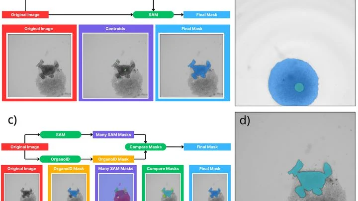
A biologist looks through a microscope at dozens of tiny cell spheres — spheroids. From these, miniature organ-like structures will later grow. The task is similar to a baker's job of checking whether the dough has risen evenly in each mold. Only here, instead of molds, there's a nutrient medium, and instead of dough, there are living cells.
A tongue tumor is like a stranger in a crowd. On the surface, they all look much alike, but beneath the surface hide very different personalities. Some tumors behave aggressively, others grow slowly. Doctors need to understand who they're dealing with before surgery.
Usually, this means taking a tissue sample — a biopsy.
A neutron spectrometer is like a blind geologist who taps on rock and guesses what's inside by the sound. It sends a beam of neutrons into a crystal and watches how they scatter. From this pattern, you can deduce how atomic magnets, spins, oscillate. But where exactly to dig and how long to listen used to be decided by a human.
Now, the TAS-AI algorithm has taken the helm.
A doctor studies a brain scan: somewhere in the tangle of tissue, a tumor is hiding. One glance picks out small spots and edges, another takes in the overall shape and location. A person combines both, but across hundreds of scans that gets tedious. A new program does the same thing automatically.
A doctor holds a stack of test results taken at different times. Each sheet on its own is just a snapshot of a single day, but what you want is a whole movie of how the disease unfolded. A neural network called Tempo learns to glue these scattered frames into a smooth reel.
The X-ray machine moves around the breast in an arc, taking several images from different angles. From these flat frames, the computer assembles a 3D picture, like putting together a mosaic from pieces. But if the mosaic pieces are crumpled or smudged, the picture turns out blurry. In tomography, the role of "crumples" is played by noise and imperfections in the imaging.
A surgeon removes a breast tumor, but how do you know no cancer cells are left at the edge? Right now, the tissue goes to a lab, and the answer comes back days later. If the margin is not clean, a second surgery is needed. The new method promises to give the answer right in the operating room, while the patient is still on the table.