How to Assemble an Aorta from Six Scans express
The body's main artery, the aorta, is not just a tube. With each heartbeat, it stretches and contracts like a rubber hose under water pressure. Seeing this motion in volume is hard: standard MRI gives only flat slices, and full 3D imaging over time requires expensive equipment and long periods lying in the scanner.
A group of researchers found a way out. They built a "library" of hundreds of typical aortic shapes, like a tailor's pattern catalog. Then they took just six flat scans from a specific patient and used an algorithm to pick the shape from the library that best matches those slices. The algorithm is like a sculptor who reconstructs an entire statue from a few contours.
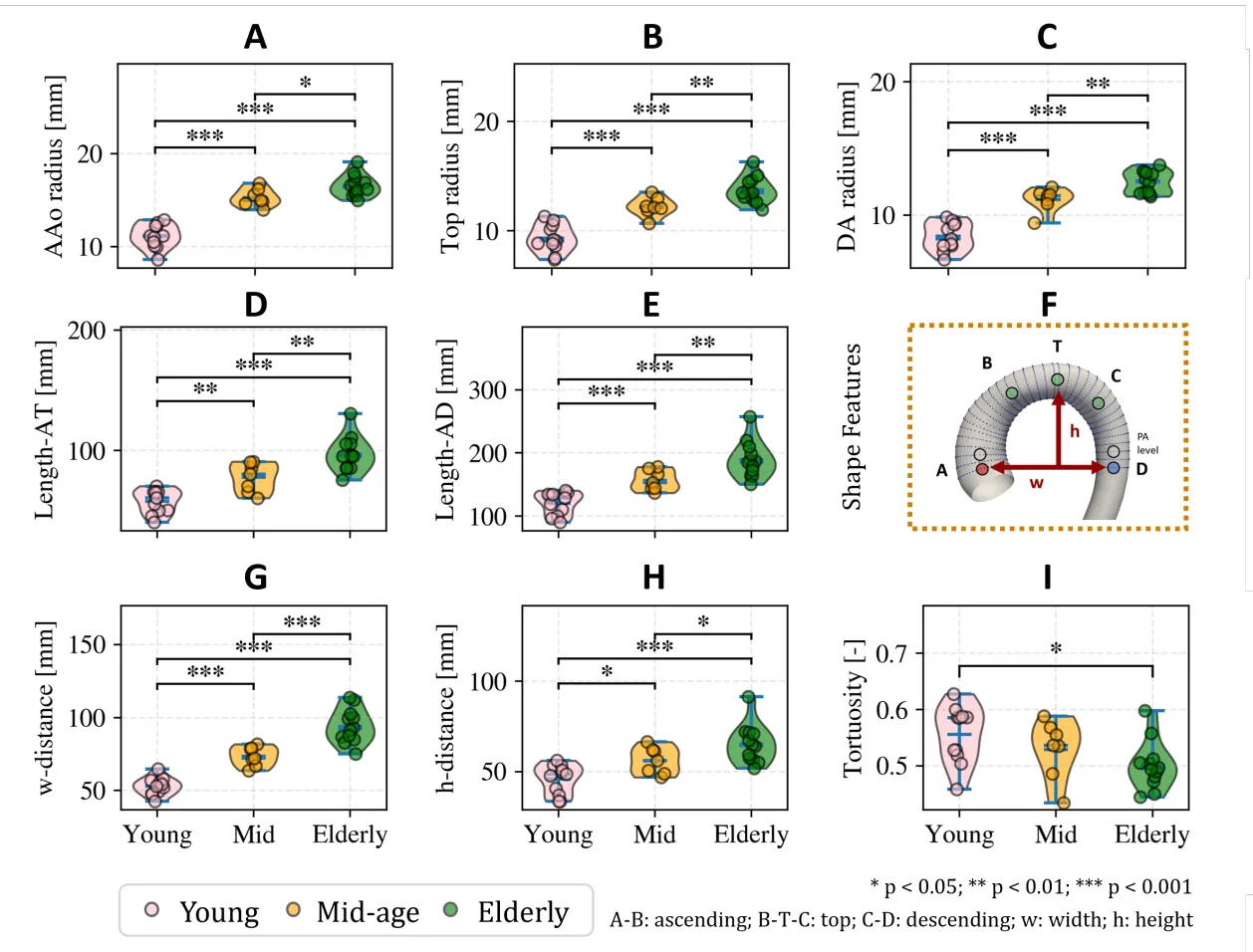
Tests on 30 volunteers and patients showed that age changes the aorta's elasticity. In young people, the wall stretches 11% with each beat; in older people, only 3%. That's the difference between a fresh rubber band and an old, cracked one.
This approach makes a 3D view of the aorta accessible in regular clinics: no new equipment needed, just smart processing of familiar scans. And that means doctors can spot dangerous changes in the main artery earlier.
The aorta is the largest blood vessel in the human body: in an adult, its diameter at the root reaches 3–4 cm, about the size of a garden hose.
In the series "The Expanse," ship doctors use portable scanners that instantly build 3D models of internal organs: this method takes a step toward such compact diagnostics.