Measuring the Invisible: Machine Learning Reads Electronic Patterns express
In twisted bilayer graphene, electrons behave like people on a crowded bus: each takes up space and gets in the way of neighbors. The strength of this "reluctance to share space" is called the Hubbard parameter U. It determines whether the material will be a conductor or an insulator, but measuring U directly is very difficult.
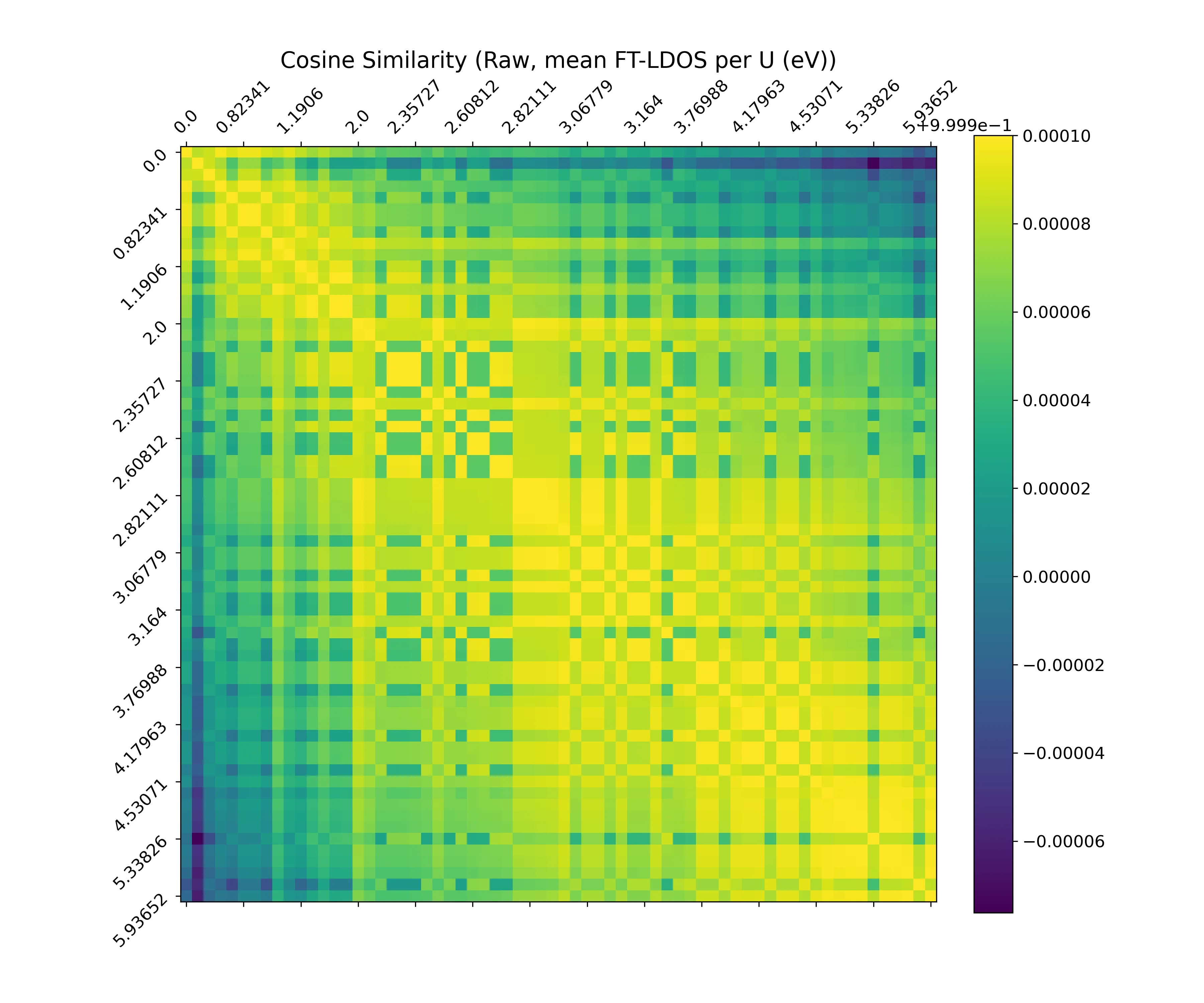
A scanning tunneling microscope (STM) takes images showing how electrons are distributed. However, for different values of U, the images look almost identical, like two photos of a crowd where people are just slightly more pressed together. The researchers trained a neural network to distinguish these subtle differences. The network looks at an image and outputs the value of U, even if the image is noisy or slightly distorted, as in real experiments.
This approach allows quick and accurate determination of the Hubbard parameter in materials where electrons interact strongly. This is important for understanding superconductivity and other unusual phenomena in layered structures.
Original abstract
as written by the authors arXiv:2602.18890
The atomistic Hubbard interaction U, representing the on-site Coulomb repulsion, serves as a pivotal parameter in theoretical models describing correlated systems, yet its precise experimental determination, especially in moire systems, remains challenging. Scanning Tunneling Microscopy (STM) provides real-space images of the local density of states (LDOS), offering rich data sets that reflect the unique electronic structure of the material. Here, we introduce a systematic methodology for extracting the Hubbard U parameter directly from these LDOS images through the application of machine learning (ML) in the case of twisted bilayer graphene in the flat-band regime. Accurate regression of U is achieved despite the extremely high visual similarity between FT-LDOS images corresponding to different interaction strengths. Subsequent data analysis further suggests a gradual interaction-dependent redistribution of spectral weight, with a possible crossover scale of order U/t ~ 1. To assess robustness beyond idealized simulations, we introduce physically motivated perturbations that emulate experimental STM imperfections and demonstrate that augmentation-based training substantially improves generalization to noisy and symmetry-broken FT-LDOS images.
A scanning tunneling microscope can "see" individual atoms by feeling the surface with a needle with precision down to fractions of a nanometer.