Laser microscope reads cell softness without a gas cell express
A laser beam passes through a cell, and the cell responds to the touch, like a taut string plucked by a finger. The frequency of this 'ring' reveals how stiff or soft the material is. That's how Brillouin microscopy works, named after the French physicist Leon Brillouin. But the method has a flaw: it's slow, because the beam scans point by point.
Speeding it up was proposed long ago — instead of a single point, send a whole line of light at once. But then too much stray light enters the instrument, drowning out the weak signal from the tissue. Previously, a gas cell was used to swallow the excess light. But the cell required a laser of strictly one wavelength — around 780 nanometers. That's like having a kitchen scale that only works with apples.
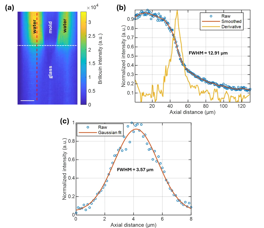
Now the gas cell is gone. In its place, two optical filters are set up in series. The first cuts off stray light, the second disperses the remaining light into colors. Together they suppress noise by 57 decibels, like the roar of a construction site outside your window fading to a whisper. The instrument became simpler and works with ordinary lasers. Tests on printed gels mimicking living tissues produced clear images.
Now such microscopes can be made cheaper and installed in ordinary labs. And crucially, they can switch to other wavelengths where scattering is stronger and the signal brighter. It's like going from a dim bulb to sunlight: you see more and faster.
The Brillouin frequency shift for water is about 5 gigahertz — a billion times higher than the note 'A' above middle C.