Biomedical spectroscopy uses the interaction of light with biological tissues to obtain diagnostic information without invasive procedures. It encompasses a range of techniques, from Raman and fluorescence spectroscopy to optical coherence tomography, that can reveal the chemical composition, structure, and function of cells and tissues in real time. This field is transforming medicine by enabling early disease detection, image-guided surgery, and personalized treatment monitoring.
History
The roots of biomedical spectroscopy go back to the 19th century with the discovery of the absorption spectrum of hemoglobin. However, the modern era began in the 1970s and 1980s with the development of fiber-optic probes and sensitive detectors, which allowed in vivo measurements. The field has since grown rapidly with the advent of new laser sources and computational methods.
How it works
Different molecules absorb, scatter, or emit light at characteristic wavelengths. By shining light on a tissue and analyzing the spectrum of the returned light, one can identify specific biomolecules, such as hemoglobin, water, lipids, or proteins, and their concentrations. Advanced techniques like diffuse optical spectroscopy use multiple source-detector distances to map these concentrations in three dimensions, while time-resolved methods can separate absorption from scattering to probe deeper into the tissue.
In practice
Biomedical spectroscopy is used in clinical settings for non-invasive blood glucose monitoring, cancer detection during endoscopy, and real-time assessment of tissue oxygenation during surgery.
Fun fact
The technique of Raman spectroscopy can identify a single molecule by its unique vibrational fingerprint, which is why it is sometimes called 'molecular fingerprinting'.
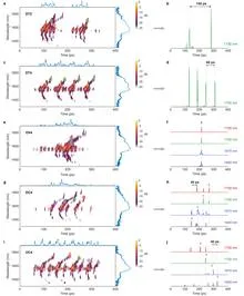
A conductor raises the baton, and the orchestra produces not just sound but a complex score: each note sounds at its own moment, at its own pitch, with the right volume. Light can also be a score, if you control its spectrum and timing. Previously, this required bulky setups that could not be reconfigured after assembly.
A thin section of eucalyptus lies under the microscope objective, and white light, passing through two polarizing filters, turns the wood into a mosaic of purple, yellow, and blue patches. These colors are not random: like letters on a map, they show the direction of the invisible cellulose threads inside the cell walls.
A cartographer receives a sheet with distances between hundreds of unnamed islands. No island is labeled, but the distances repeat, forming triangles and quadrilaterals. From these repetitions, the cartographer reconstructs the map of the archipelago, not knowing which island is which. This is roughly how the new work in molecular spectroscopy works.
In a dark room, a sunbeam passes through a pinprick in cardboard, and an upside-down image of the street appears on the wall. A pinhole camera needs no lenses: the tiny hole itself selects the rays coming from each point of the scene. Scientists at the ESRF applied this principle not to light but to X-rays, and made the camera see not shape but chemical composition.
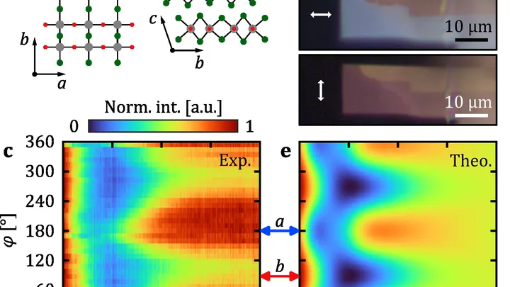
Light born when a fast electron enters a material is usually assumed to be the same in all directions. But in crystals where optical properties depend on direction, that's not the case. Run your hand over velvet: one way it's smooth and shiny, the other — dark and rough. Similarly, a crystal responds differently to light depending on how it's oriented.
A doctor looks at a screen and sees two images of the same part of the body. One shows where blood is rich in oxygen, the other shows how fast it rushes through the finest vessels. Previously, these images were made by different devices, and combining them was difficult. Now engineers have built a device that takes both images simultaneously.
A drop of blood from a fingertip lands on the white membrane of a cartridge. Twenty minutes later, the phone shows whether Lyme disease is present or not. No test tube, no centrifuge, no waiting for days.
Usually, for such a test, blood is first spun in a centrifuge to separate the liquid part — the plasma.
Light passing through a glass of murky water dims — some of it is absorbed. That's how many liquid-analysis instruments work: they measure how much light was lost and infer the composition from that. But if the liquid is heterogeneous, like soup with chunks, a single number isn't enough: you need to know where exactly more absorption happened.
A stack of playing cards lies still. You can slide the top card with your finger: that's crude intervention. Or you can blow so that the whole stack momentarily changes shape, but the cards don't scatter. That's roughly how light, in a new experiment, rearranges a crystal of tin selenide (SnSe).
Usually, to change the structure of a solid, you heat it or pass a current through it.
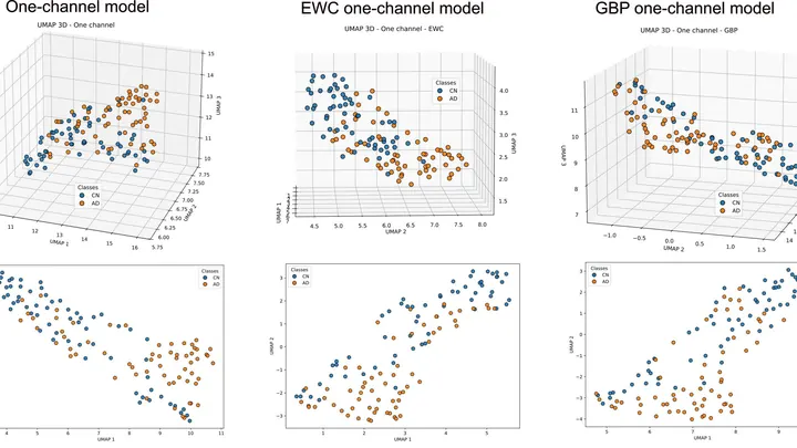
A doctor spends years learning to recognize diseases from brain scans. First, they excel at spotting signs of aging, then they master tumors, and suddenly they start confusing what they knew before. Many neural networks behave much the same way: new knowledge overwrites the old.
A bell hidden in a thick box can be made to ring without opening the lid. Just send a sound wave through the walls, and the clapper will tremble. That's roughly how transcranial ultrasound works: sound passes through the skull bones and makes neurons deep in the brain 'ring'—generating electrical impulses.
Radiation therapy is a precision strike on a tumor, but every shot must be calibrated to within a millimeter and a fraction of a second. The problem is that existing dosimeters are either bulky, or they distort the beam themselves, or they require pauses for readout.
A single-pixel camera works like an artist creating a picture through a stencil with many holes. In each frame, only a portion of the light, modulated by a mask, a spatial light modulator, reaches the single sensor, and then the computer reconstructs the full image by assembling this mosaic with computational algorithms.
Medical scans are maps of tissue density. The brightness of each pixel reveals what’s there, much like photometry measures light intensity. But conventional algorithms analyze pixels in isolation, producing jagged organ boundaries.
A new approach compels the program to prioritize smoothness.
Tomographs don't produce ready-made pictures. They collect numerous 'shadows' — projections of innards from different angles or using various signals — and mathematically reconstruct a 3D picture from them.
Each positron annihilation is like the birth of a pair of dancers, connected by an invisible thread of polarization. Two gamma quanta fly apart, but their states remain entangled: measuring one instantly determines the other — as if one partner senses every movement of the other, no matter the distance.
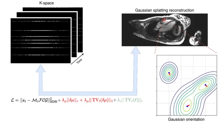
A photographer shoots a runner with a long exposure and gets a blurry silhouette. That's roughly how a standard MRI scanner "sees" moving organs: the heart contracts, the lungs breathe, and the image smears. To avoid this, patients are asked to freeze and hold their breath. But for a child or someone short of breath, that's nearly impossible.
The method called GRASP tackles the problem differently.
When you need to see inside the brain, you usually lie in a tight tube of an MRI scanner or get zapped with X-rays. Both methods are costly, bulky, and not always available.
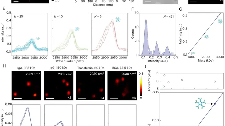
A drop of blood from a vein falls under three beams, and each one drags its own clues into the light. The infrared beam makes molecules vibrate — from that trembling, you learn how much sugar and fat is in the sample. A green laser scatters off proteins and lipids, shifting color — revealing the remodeling of cell membranes.
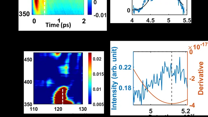
A short laser pulse hits an iodine molecule, and its atoms start to vibrate like a ball on a spring. Scientists track these vibrations with a second, broadband pulse that captures a spectrum at different moments in time.
It turns out that vibrations in the excited B state quickly shift in frequency and fade out, and then vibrations appear in the A state.
A candle flame flickers in a draft but doesn't go out — the tongue of fire briefly changes shape and then returns. Optical skyrmions behave much the same way — tiny vortices in a light beam, reminiscent of swirling eddies. Until recently, they were created only in the perfect, smooth light of a laser.
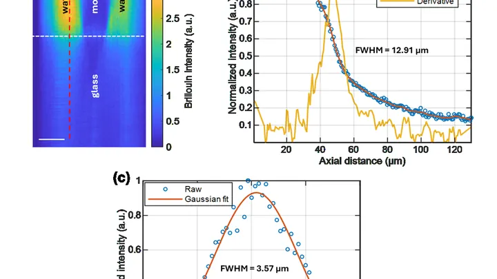
A laser beam passes through a cell, and the cell responds to the touch, like a taut string plucked by a finger. The frequency of this 'ring' reveals how stiff or soft the material is. That's how Brillouin microscopy works, named after the French physicist Leon Brillouin.
Your finger is a living sensor: beneath the skin, arteries pulse in time with your heart. Traditional blood pressure cuffs squeeze your arm and give a single number, but this ring uses eight electrodes to pass a weak current through the tissue and map blood flow.
Inside every cell, there are tiny power stations — mitochondria. They burn nutrients and store energy. Two helper molecules, NADH and FAD, oversee this process. Previously, they were detected by their faint glow, but it was dim, and signals from different molecules overlapped.
The new microscope works differently.
A drummer stays silent, but from the pauses you can tell where he didn’t hit. Similarly, physicists built an image by catching not light, but moments of absolute darkness. The picture emerged from silence.
In the experiment, they shone light from an ordinary lamp onto the object.
Inside every cell, there's a network of thin tubes — microtubules. They act as both scaffolding and rails: they hold the cell's shape and transport cargo. These tubes are built from the protein tubulin and naturally glow faintly under ultraviolet light. Scientists wondered: can we make this glow stronger or weaker on demand?
The key to control is the amino acid tryptophan.
Atoms in a beam travel at different speeds, like runners on a track. A regular laser pulse only pushes those moving at one speed, but the new one acts like a coach giving commands to the whole group at once. This approach involves more atoms in the measurement.
A pathologist stains a tissue section with hematoxylin and eosin — cells turn blue and pink, and under the microscope you can see their shape and arrangement. But which genes are active in each cell cannot be read from such an image. That requires a separate, expensive assay that reveals the activity of thousands of genes at once, but it is rarely performed.
A mole on your skin looks like a mosaic of colored pieces. Each piece is a small patch of the image where color and texture barely change. Such pieces are called superpixels. If you connect them with lines, you get a network that looks like a subway map: stations are the pieces, and lines are the connections between them. The weight of each line shows how strongly two pieces are related.
A conventional light microscope hits a fundamental limit: it cannot distinguish two points closer than 200 nanometers — roughly the thickness of a soap bubble divided by a hundred. To see deeper, scientists have made molecules in the sample blink or fade, but these tricks are slow and harmful to living cells.
Infrared light is invisible to the eye, but it's everywhere: from TV remotes to the heat of the human body. Catching it requires special materials. Conventional detectors are bulky, rigid, and not easily tuned to specific wavelengths.
A quantum dot is like a tiny lamp that turns on and off at the command of a laser, emitting exactly one photon at a time. But this light has a quirk: the photons don't interact with each other; they fly independently, like random drops from a faucet.
A protein in a cell is like a tightly coiled spring. Its shape determines what it does, but seeing that shape in a single molecule without disturbing it was nearly impossible. The new microscope works differently: it sends a short laser pulse that slightly heats the molecule.
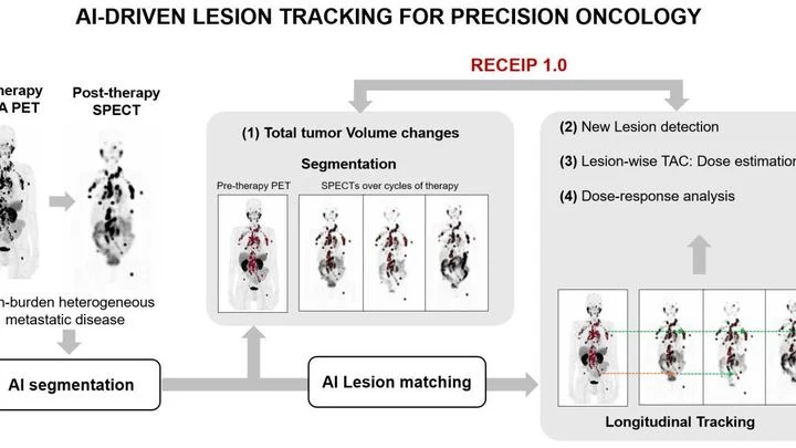
When treating prostate cancer, doctors often see not one tumor but many lesions in bones and lymph nodes. Each lesion can respond to therapy differently: one shrinks, another stays the same, a third grows. Previously, the disease was assessed as a whole, like judging the weather by the average temperature over a year.
In a hospital, blood from a finger prick usually goes to the lab, and the result comes back the next day. The new method promises to cut that wait to a few minutes. Instead of catching light reflected off a sample, it reads the electrical voltage that arises at the boundary between two media, like the faint tingle you feel when you touch a battery with wet fingers.
An MRI scanner is like a camera that photographs your insides, but very slowly. The patient has to lie still for several minutes while the heart keeps beating. If the person moves or the rhythm goes off, the image blurs. Doctors then have to repeat the scan, wasting time and nerves.
Entangled photons are like two couriers marching in lockstep: their quantum states, particularly polarization, are synchronized so that measuring one instantly determines the state of the other, even at a distance. In a material, this synchronization gets disrupted.
To avoid losing a fast soccer player in the frame, a drone doesn't need a bright beacon — recognizing the silhouette is enough. Similarly, a neural network now tracks microbes: the algorithm analyzes an ordinary microscope image and instantly finds a bacterium in a drop of water.
A finger is illuminated with a laser beam, and some of the light returns after traveling inside. That's how Raman spectroscopy works: molecules in bone and skin alter the color of the reflected beam differently, leaving their chemical fingerprints. But skin, muscle, and fat are noisy: their signal drowns out the bone's, like a crowd's chatter overpowering a single singer.
Strike a tuning fork, and it rings with a single pure note. But if the fork is trembling and people are chattering nearby, the sound won't be perfect. Same with molecules: each one absorbs light of a specific color, but due to its own vibrations and the jostling of neighbors, that color gets smeared out.
The authors of the paper figured out how to show this to students without complex formulas.