How MRI Peers Inside Nerve Fibers express
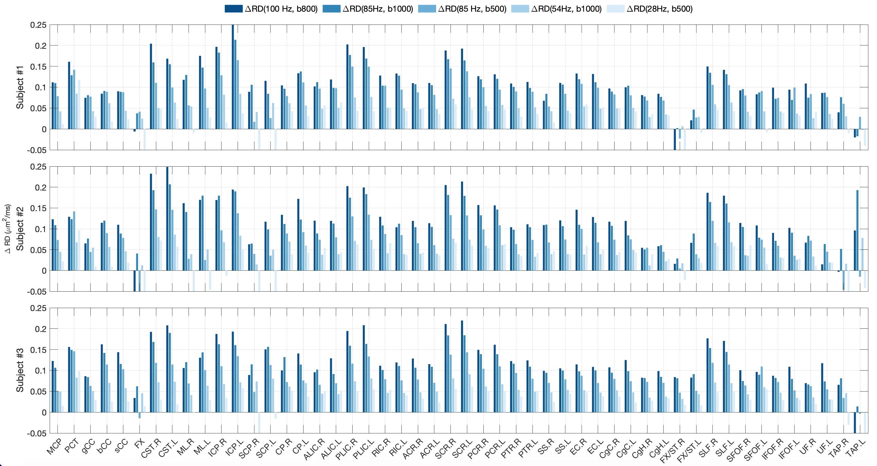
Water inside nerve fibers doesn't move like it does in a glass: it constantly bumps into the walls of axons (the long projections of nerve cells). If you make the pause between MRI radio pulses very short, water doesn't have time to travel far, and its movement reflects the tightness of the surrounding space.
In the new study on living humans, they compared water mobility across fibers at short and long observation times. The difference was especially striking in the corticospinal tract — the main highway through which the brain commands muscles. That's where axons are thickest, and water takes longer to travel from one wall to the other.
Previously, such differences could only be seen in tissue samples under a microscope. Now MRI with oscillating gradients does the same without a single cut. The method promises to become a tool for tracking axon damage in multiple sclerosis and other diseases — long before symptoms appear.
Original abstract
as written by the authors arXiv:2607.25749
Diffusion time-dependence, defined as variations in diffusivity and/or diffusional kurtosis with diffusion time, has emerged as a valuable non-invasive imaging marker for characterizing tissue microstructural features, such as cell size, density, packing disorder, and membrane permeability. In white matter, diffusion time-dependent changes between the short diffusion time and long diffusion time in radial diffusivity (RD), defined as the diffusivity perpendicular to fiber tracts, were demonstrated to correlate strongly with mean axon diameter in ex vivo spinal cord tissues, and to reveal demyelination in mouse corpus callosum. Despite their potential to non-invasively unveil neuronal microstructures to improve the assessment and targeted therapy of neurological diseases, these novel image contrasts obtained at short diffusion times using oscillating gradient spin echo (OGSE) have only recently become feasible for human in vivo studies with high-performance gradient MRI systems. In this preliminary study, we characterized time-dependent RD with OGSE encoding in the human brain in vivo. The change in radial diffusivity between short diffusion time and long diffusion time (delta_RD) consistently exhibited high values in the corticospinal tract, indicating high sensitivity of delta_RD to large axon diameter in human brains. Imaging at a high OGSE frequency of 100 Hz and a moderate b-value of 800 s/mm2 produced the highest delta_RD in the corticospinal tract. This study established a baseline for future investigations of neuronal microstructural alterations in neurological disorders and diseases.
In the corticospinal tract, axons can reach a meter in length: they are the longest cells in the human body.
In 'The Matrix,' the brain was plugged in directly, but even there they couldn't measure axon thickness without cutting open.