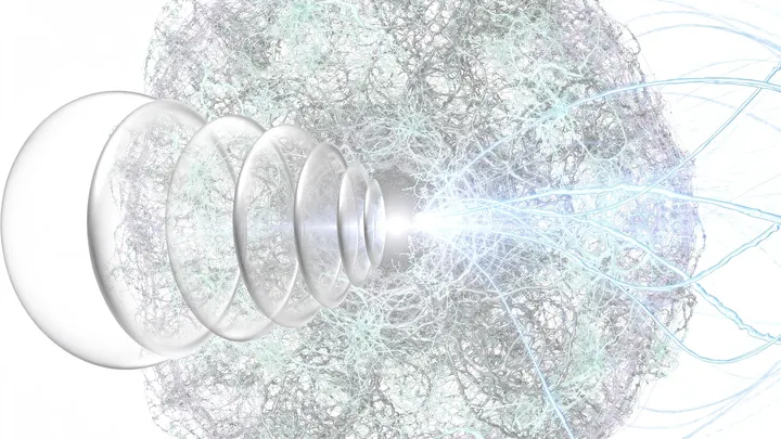
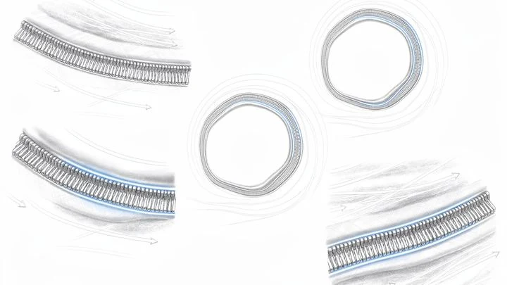
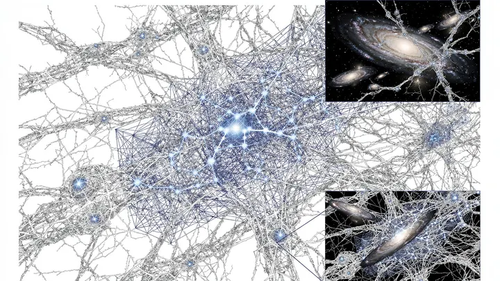
Diffusion MRI is a magnetic resonance imaging technique that measures the random motion (diffusion) of water molecules in biological tissues. Because water diffusion is hindered by cellular structures — membranes, organelles, and fibers — the measured diffusion pattern encodes information about tissue microstructure at a scale much smaller than the imaging resolution. This makes diffusion MRI a powerful tool for non-invasively probing the architecture of organs like the brain, heart, and muscles.
History
Diffusion MRI was pioneered in the mid-1980s by Denis Le Bihan and colleagues, who developed the concept of diffusion-weighted imaging (DWI). The diffusion tensor model was introduced in the early 1990s by Peter Basser and collaborators, enabling the mapping of fiber orientations.
How it works
The technique uses magnetic field gradients to sensitize the MRI signal to molecular displacement. In a typical diffusion-weighted sequence, two strong gradient pulses are applied: the first dephases the spins, and the second rephases them. If water molecules have moved between the pulses, the rephasing is incomplete, leading to signal attenuation. The amount of attenuation is related to the diffusion coefficient along the gradient direction. By applying gradients in multiple directions, one can reconstruct a diffusion tensor that describes the magnitude and directionality of water motion — revealing, for example, the orientation of white matter fiber tracts in the brain.
In practice
Diffusion MRI is widely used in clinical neurology for diagnosing stroke, in neurosurgery for surgical planning, and in research for studying brain connectivity. It is also applied to other tissues, such as muscle and cardiac imaging, and is being explored for cancer characterization.
Fun fact
The technique can reveal the internal structure of cells without a microscope, as highlighted in the article 'Как разглядеть, что внутри клетки'.
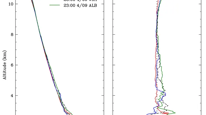
Every second, thousands of passenger aircraft in the sky transmit radio signals: that's how air traffic controllers track where each flight is. These signals don't travel in a straight line: air of varying humidity bends them like a lens. If you can measure that bending, you can work out where the air is drier and where it's more humid.
A doctor looks at a screen and sees two images of the same part of the body. One shows where blood is rich in oxygen, the other shows how fast it rushes through the finest vessels. Previously, these images were made by different devices, and combining them was difficult. Now engineers have built a device that takes both images simultaneously.
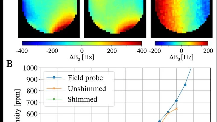
An MRI scanner is a big magnet that aligns hydrogen atoms in the body and then picks up their response signal. Such machines usually take up an entire room and cost as much as a nice house. Engineers from several labs decided to build a scanner that fits in a suitcase and released all the plans as open source.
A sponge slowly watered from a watering can gets soaked all the way through. But if you dump the same bucket of water on it all at once, the middle gets wet while the edges stay almost dry. That's roughly how a material with nonlinear absorption behaves: the more intense the flux, the more it gets stuck at the point of impact.
Light passing through a glass of murky water dims — some of it is absorbed. That's how many liquid-analysis instruments work: they measure how much light was lost and infer the composition from that. But if the liquid is heterogeneous, like soup with chunks, a single number isn't enough: you need to know where exactly more absorption happened.
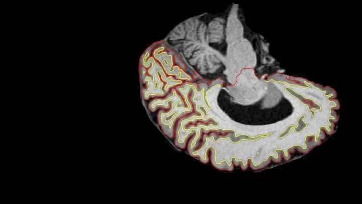
Two scans of the same brain taken at different times are like photographs of the same house before and after renovation. To see what changed, you overlay them and look at the differences. But an MRI scanner is not a perfect camera: it adds its own distortions, as if the photo were taken through slightly warped glass.
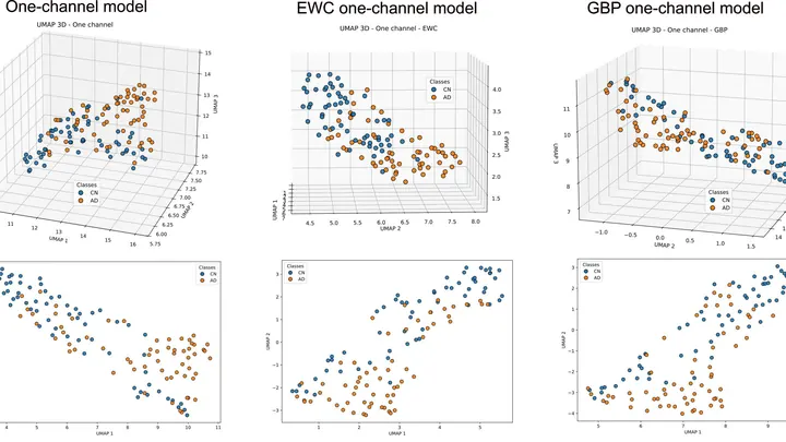
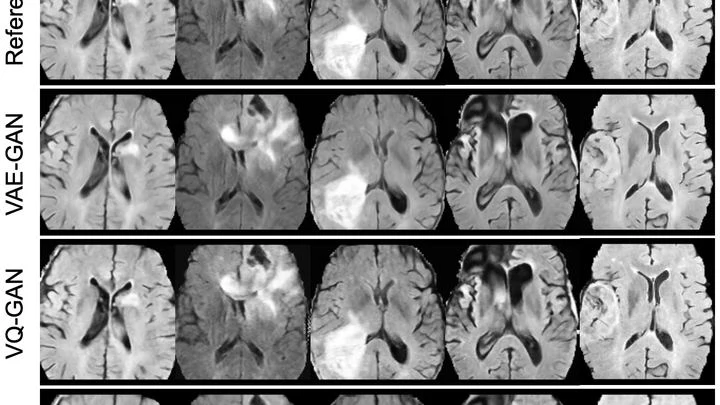
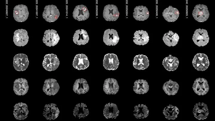
A doctor spends years learning to recognize diseases from brain scans. First, they excel at spotting signs of aging, then they master tumors, and suddenly they start confusing what they knew before. Many neural networks behave much the same way: new knowledge overwrites the old.
Radiation therapy is a precision strike on a tumor, but every shot must be calibrated to within a millimeter and a fraction of a second. The problem is that existing dosimeters are either bulky, or they distort the beam themselves, or they require pauses for readout.
A neural network predicts the future of a system — say, how plasma will swirl in a magnetic field or how a face in a video will change. Errors are inevitable, and the further it looks ahead, the more they accumulate. Checking against the correct answer during operation is impossible: it simply doesn't exist.
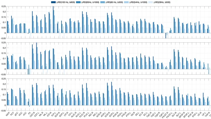
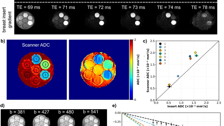
Water inside nerve fibers doesn't move like it does in a glass: it constantly bumps into the walls of axons (the long projections of nerve cells). If you make the pause between MRI radio pulses very short, water doesn't have time to travel far, and its movement reflects the tightness of the surrounding space.
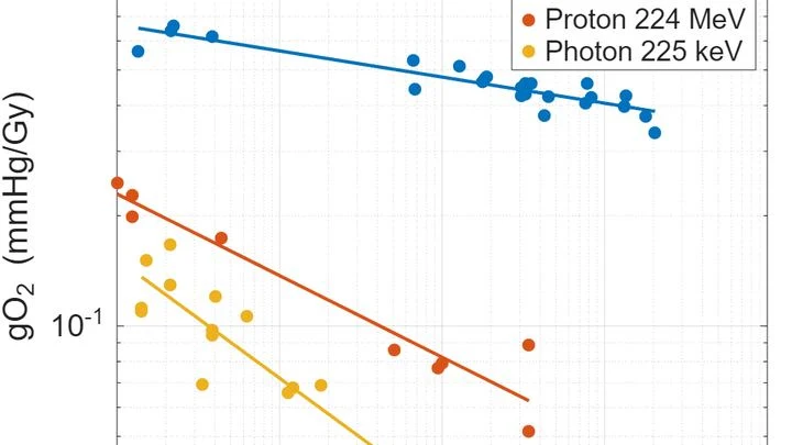
A camera flash that fires in a thousandth of a second doesn't heat the subject — unlike a regular flash that lasts a full second. A similar trick has been found in radiotherapy: when a radiation dose is delivered in milliseconds instead of minutes, healthy tissues suffer less. This is called the FLASH effect.
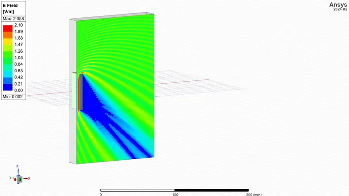
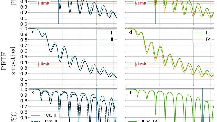
When a radio wave encounters a building in its path, it doesn't just reflect or get absorbed: it bends around it, much like ocean waves flowing around a rock, leaving calm water behind. In radiophysics, this phenomenon creates a radio shadow — a complex pattern of zones where the signal is amplified or attenuated, which scientists call a radio portrait.
The data from a single CT scan is comparable to several feature-length films. TCAM-Diff bypasses this bottleneck: the program compresses the 3D scene into three shadow projections — like a sculptor reconstructing a solid statue from silhouettes on three walls.
Prostate MRI scans often look like photos taken through a dirty window: noise, blurred edges, curvature from field imperfections. Scientists checked what happens if you 'wash' that window: remove the excess, correct the distortions, and accurately align different types of scans.
In the brain's white matter, the signal travels along axons — long neuronal processes wrapped in an insulating myelin sheath. Water fills the space inside axons and outside, in the narrow gaps between fibers.
Fast radio bursts — millisecond radio pulses arriving from distant galaxies — long remained a puzzle. Their colossal energy, compressed into instants, pointed to cataclysms on neutron stars, perhaps pulsars or magnetars.
Membraneless organelles, tiny factories inside the cell, defy conventional rules. Take the nucleolus: here ribosomal RNA is synthesized — long chains woven from carbon sugars, rich in oxygen phosphate groups, and stitched with hydrogen bonds, suspended in the watery environment of the nucleus.
Light from distant galaxies travels to us for billions of years, but along the way it gets bent and twisted by the invisible web of dark matter. This effect, predicted by Albert Einstein within general relativity, is called gravitational lensing.
An ordinary microscope is powerless when you need to examine a single virus in detail. It's too small. But there's a tool that fires flashes billions of times brighter than sunlight — an X-ray free-electron laser. It's designed to 'photograph' a particle before the beam vaporizes it.
In a small Petri dish sits a droplet of liquid the size of a pinhead. An ordinary scanner wouldn't even notice it: the signal is too weak. But bring a tiny magnetic field sensor close to the droplet, and the picture changes.
Physicists have achieved three-dimensional Anderson localization of electromagnetic waves — a phenomenon where waves become permanently entangled in a random labyrinth of metal particles and cease to propagate.
A flashlight spreads light in a broad beam, while a laser cuts a narrow path and picks out the finest details. In magnetic resonance imaging (MRI), the gradient, a rapid change in the magnetic field along the body, plays the role of that laser.
To peek inside a muscle, it is illuminated with radio waves in a strong magnetic field: that's how MRI works. Water in cells and water in blood respond differently, but a standard scanner doesn't tell them apart: it produces an averaged image where the signal from capillaries is superimposed on the signal from muscle fibers.
A drop of ink in a glass of water spreads quickly and evenly. But if the water is squeezed between tightly packed spheres, the ink cloud crawls slowly and erratically: it dodges obstacles and gets stuck in narrow necks. Water molecules in living tissue move in much the same way.
Imagine a water park with many pools connected by narrow passages. Visitors, water molecules, splash around in each pool, but occasionally move to the next one. In a brain tumor, these "passages" are protein channels in cell membranes. Their abundance determines how quickly water moves between cells.
A distant lighthouse flashes for an instant brighter than an entire galaxy. This is a fast radio burst — a mysterious signal from deep space. Some repeat, and astronomers have noticed a peculiarity: these bursts show rapid changes in polarization — a property indicating how the radio wave is twisted in space.
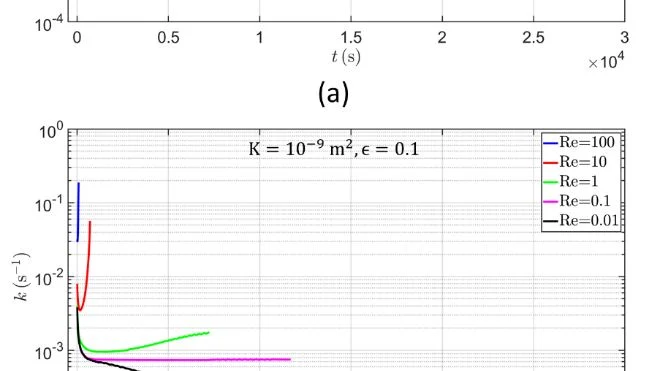
A porous implant filled with a drug works like a sponge that slowly releases the substance into the surrounding fluid. But if the fluid flows quickly, it washes out the drug differently than in a calm environment. The authors computed on a computer how different flow rates and properties of the porous structure affect drug release.
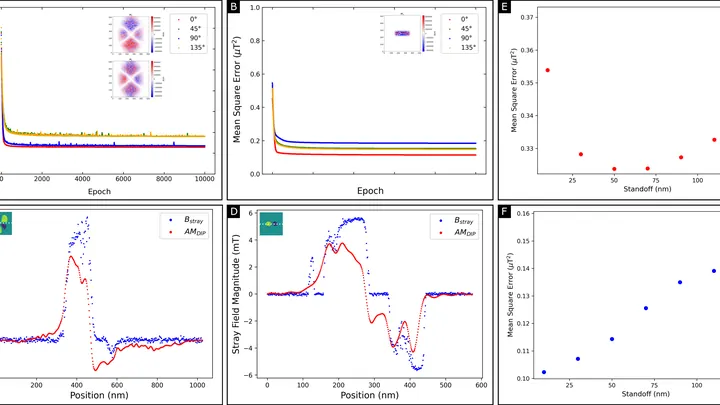
A magnetic film just tens of atoms thick stores information in tiny magnetic vortices. But you can't look inside and see these vortices directly: the instrument only senses the weak magnetic field around the film. Reconstructing the magnetization pattern from such a field is like guessing the shape of an invisible relief from its shadow on a wall.
The brain is a fortress. It's protected by a dense layer of cells that only lets in what it needs. Drugs can't get through this barrier, which is why many brain diseases are so hard to treat.
Scientists have found a way to crack the fortress gates. They built a device that sends focused ultrasound into the brain. Tiny gas bubbles are injected into the blood.
A surgeon removes a breast tumor, but how do you know no cancer cells are left at the edge? Right now, the tissue goes to a lab, and the answer comes back days later. If the margin is not clean, a second surgery is needed. The new method promises to give the answer right in the operating room, while the patient is still on the table.
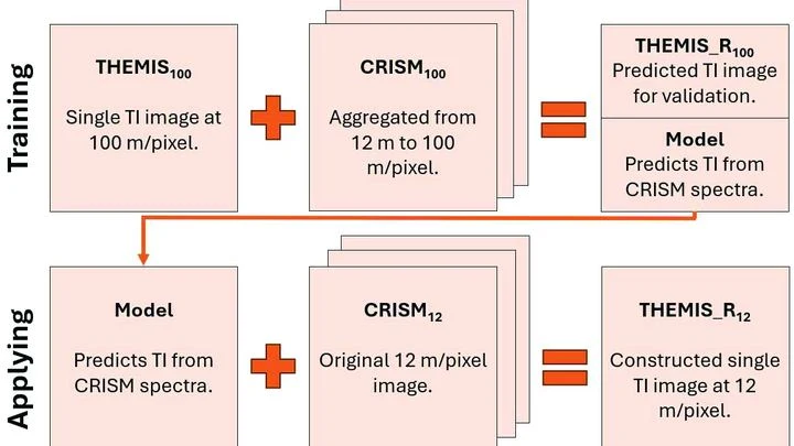
Warm rock and cold sand cool at different rates at night. An instrument orbiting Mars measures this heat, revealing whether the surface is loose dust, a dense crust, or solid rock. But thermal images have a resolution of 100 meters per point: an entire football stadium fits into a single pixel. Fine details (the beds of ancient streams, the edges of lava flows) are simply invisible.
A thin film for a solar cell or display is like a dense crowd in a square: each person faces a different direction, and whether light passes through the crowd or gets stuck depends on which way everyone is looking. In organic electronics, molecules play the role of passersby. Until now, instruments could only measure the crowd's overall direction — the average head tilt and its spread.
Your finger is a living sensor: beneath the skin, arteries pulse in time with your heart. Traditional blood pressure cuffs squeeze your arm and give a single number, but this ring uses eight electrodes to pass a weak current through the tissue and map blood flow.
Imagine a city where streets are not straight but winding, with dead ends and hidden passages. A bike courier will take much longer to deliver a package than in a city with a regular grid of roads. Similarly, molecules inside living tissue: their path depends on how "tangled" the tissue structure is.
Scientists built a model that describes the movement of molecules in such a "tangled" environment.
In a disk of gas and dust around a young star, dust particles behave like sand grains in a stream of water: the gas flows faster, the dust lags behind, and friction arises between them. This friction can rock the magnetic waves that run through the disk. The galaxy is full of such disks: it is precisely in them that planets are born.
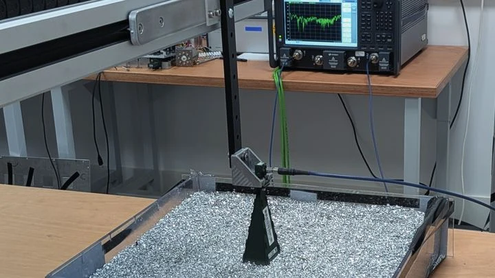
An ordinary camera captures light reflected from objects and builds a picture from it. But if the object is hidden behind an obstacle or does not reflect light, radio waves come to the rescue: they pass through walls and fog, and a computer reconstructs the image from scattered signals.
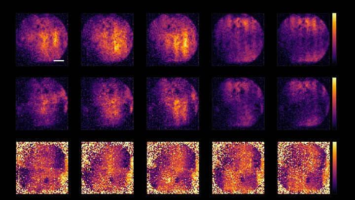
Biologists often tag cells with glowing molecules — it's like searching for a dim light in the smoke. The light quickly fades, and the smoke creates interference.
A conventional light microscope hits a fundamental limit: it cannot distinguish two points closer than 200 nanometers — roughly the thickness of a soap bubble divided by a hundred. To see deeper, scientists have made molecules in the sample blink or fade, but these tricks are slow and harmful to living cells.
On the surface of a thin film of bismuth iron oxide, you can see neat stripes, like furrows in a plowed field. What lies beneath them has remained a mystery until now.
In a hospital, blood from a finger prick usually goes to the lab, and the result comes back the next day. The new method promises to cut that wait to a few minutes. Instead of catching light reflected off a sample, it reads the electrical voltage that arises at the boundary between two media, like the faint tingle you feel when you touch a battery with wet fingers.