Magnetic resonance imaging (MRI) is a non-invasive medical imaging technique that produces detailed three-dimensional images of the body's internal structures, particularly soft tissues like muscles, the brain, and the heart. Unlike X-rays or CT scans, it does not use ionizing radiation, making it safer for repeated use. MRI is essential for diagnosing a wide range of conditions, from torn ligaments and spinal cord injuries to brain tumors and cardiovascular disease.
History
The principles of nuclear magnetic resonance were discovered in the 1930s by Isidor Rabi, and the medical application was developed by Paul Lauterbur and Peter Mansfield in the 1970s, who shared the 2003 Nobel Prize in Physiology or Medicine for their work.
How it works
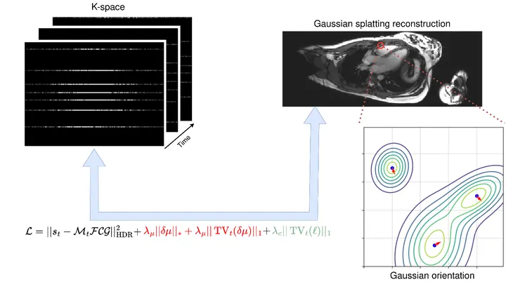
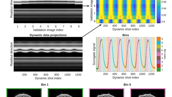
The patient lies inside a powerful superconducting magnet that aligns the magnetic moments of hydrogen nuclei (protons) in water and fat molecules. A radio-frequency pulse is then applied, knocking these protons out of alignment. As they relax back to their original state, they emit radio signals that are detected by receiver coils. By applying magnetic field gradients, the signals are spatially encoded, and a computer reconstructs them into a high-resolution image. Advanced techniques, such as real-time imaging of the beating heart, use sophisticated algorithms to accelerate data acquisition.
In practice
MRI is used in hospitals worldwide for diagnosing neurological disorders, musculoskeletal injuries, and cardiac function, as well as in research to study brain activity and muscle dynamics in real time.
Fun fact
The magnetic field of an MRI machine is so strong that it can pull a wheelchair across a room, which is why strict safety screening is required before entry.
Light knocks electrons out of a crystal, and they fly toward the detector like arrows toward a target. From their trajectories, physicists reconstruct how electrons behaved inside the material: with what energy and in which direction they moved. But switch on a magnetic field, and the trajectories bend. The arrows start to curve, the target turns into a warped mirror, and the picture blurs.
A radiologist looks at an abdominal scan and sees not just organs but their spatial relationships: which kidney is larger, where the tumor lies relative to a vessel. It's like reading a map, where it's not enough to know where a city usually stands; you also need to measure the distance between two points on the ground.
A doctor looks at a screen and sees two images of the same part of the body. One shows where blood is rich in oxygen, the other shows how fast it rushes through the finest vessels. Previously, these images were made by different devices, and combining them was difficult. Now engineers have built a device that takes both images simultaneously.
A bowl of jelly trembles but holds its shape when given a gentle shake. Thick syrup, in response to the same motion, sloshes and spreads. A healthy liver is like jelly, but an inflamed one at low frequencies becomes more like syrup.
Researchers built a device that shakes the liver at twelve different frequencies — from five to eighty oscillations per second.
An MRI scanner is a big magnet that aligns hydrogen atoms in the body and then picks up their response signal. Such machines usually take up an entire room and cost as much as a nice house. Engineers from several labs decided to build a scanner that fits in a suitcase and released all the plans as open source.
A doctor spends years learning to recognize diseases from brain scans. First, they excel at spotting signs of aging, then they master tumors, and suddenly they start confusing what they knew before. Many neural networks behave much the same way: new knowledge overwrites the old.
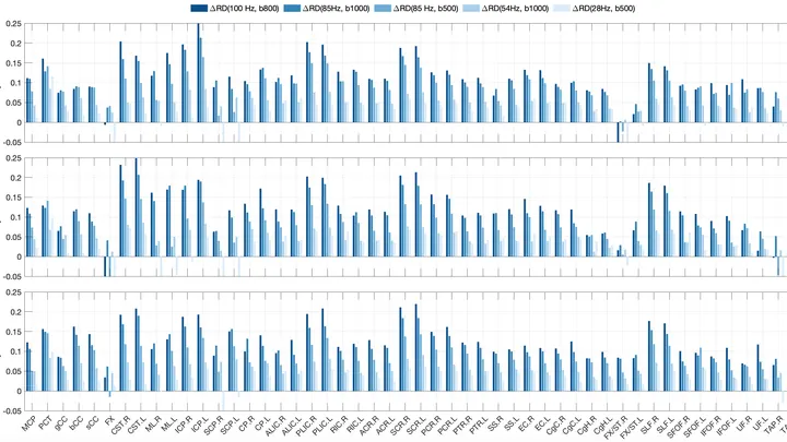
Water inside nerve fibers doesn't move like it does in a glass: it constantly bumps into the walls of axons (the long projections of nerve cells). If you make the pause between MRI radio pulses very short, water doesn't have time to travel far, and its movement reflects the tightness of the surrounding space.
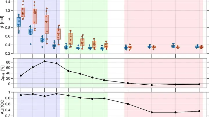
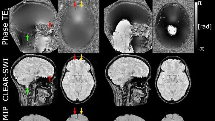
The powerful magnet of an MRI scanner induces weak electric currents in tissues. Their phase behaves like a compass needle trembling with every gust of wind — the slightest disturbance turns the map into a ripple, where instead of small vessels or iron deposits, all you see is noise.
A photographer shoots a runner with a long exposure and gets a blurry silhouette. That's roughly how a standard MRI scanner "sees" moving organs: the heart contracts, the lungs breathe, and the image smears. To avoid this, patients are asked to freeze and hold their breath. But for a child or someone short of breath, that's nearly impossible.
The method called GRASP tackles the problem differently.
Your arm lifts a cup from the shelf — a familiar motion you don't notice until muscles tire. Then the trajectory changes: the elbow shifts slightly, the hand trembles. A doctor usually catches these changes by hand, feeling the patient's muscles.
An X-ray tomograph is like a carpenter who examines a log from all sides to see where the void is inside. It takes dozens of flat shadow images, rotating the source and detector around the object. Then a 'reconstruction' program glues these shadows into a 3D model. The quickest reconstructor is the FBP algorithm (filtered back projection).
To peek inside a muscle, it is illuminated with radio waves in a strong magnetic field: that's how MRI works. Water in cells and water in blood respond differently, but a standard scanner doesn't tell them apart: it produces an averaged image where the signal from capillaries is superimposed on the signal from muscle fibers.
A figure skater glides across the ice, gradually moving toward the center. Their trajectory isn't a perfect spiral: sometimes they catch the rhythm of the music and add an extra turn, slightly changing their speed. That's roughly how a small black hole behaves when it falls into a supermassive one.
Your finger is a living sensor: beneath the skin, arteries pulse in time with your heart. Traditional blood pressure cuffs squeeze your arm and give a single number, but this ring uses eight electrodes to pass a weak current through the tissue and map blood flow.
An ordinary camera captures light reflected from objects and builds a picture from it. But if the object is hidden behind an obstacle or does not reflect light, radio waves come to the rescue: they pass through walls and fog, and a computer reconstructs the image from scattered signals.
A weak magnet in an MRI machine is like a dim bulb in a dark room: you get a picture, but details drown in grain. These scanners operate at fields below 0.1 tesla (a typical hospital machine runs at 3 tesla, 30 times stronger), yet they're lightweight, inexpensive, and don't require a special room. The catch: noise eats away at quality.
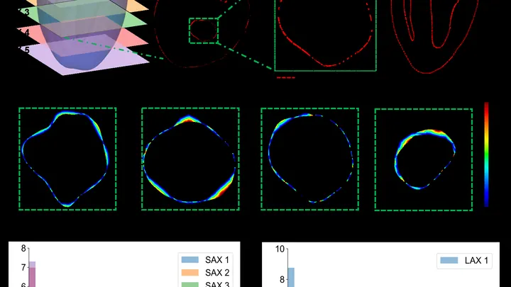
The heart is not a perfect sphere; its shape changes with every beat. To build an accurate 3D copy for a computer model, doctors typically take dozens of MRI slices. But that's like assembling a watermelon from thin slices: the fewer slices, the more guesswork.
A new approach teaches a neural network to understand the heart's shape from sparse contours.
An MRI scanner is like a camera that photographs your insides, but very slowly. The patient has to lie still for several minutes while the heart keeps beating. If the person moves or the rhythm goes off, the image blurs. Doctors then have to repeat the scan, wasting time and nerves.
A finger is illuminated with a laser beam, and some of the light returns after traveling inside. That's how Raman spectroscopy works: molecules in bone and skin alter the color of the reflected beam differently, leaving their chemical fingerprints. But skin, muscle, and fat are noisy: their signal drowns out the bone's, like a crowd's chatter overpowering a single singer.
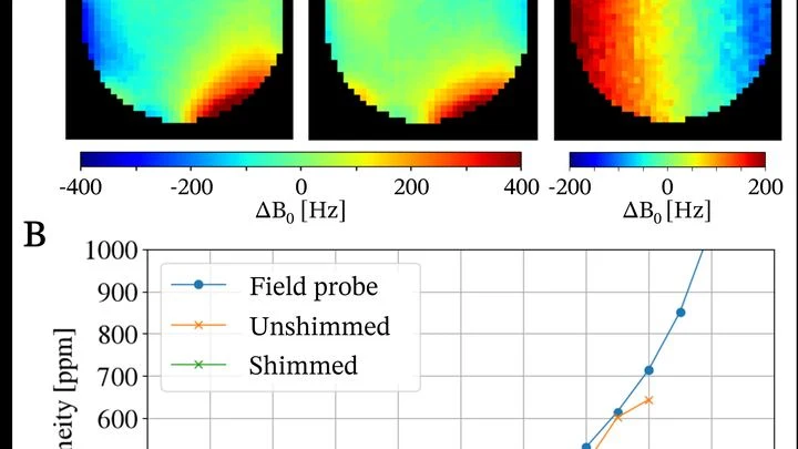
A standard MRI machine is a huge magnet that you can't move, plus a powerful computer that turns signals from the body into a picture. In recent years, portable scanners with weaker fields have appeared: they are light, cheap, and can work in a field hospital or an ambulance.
The heart doesn't contract all at once but as a wave: an electrical signal travels through the muscle, and behind it, like ripples spreading on water, a contraction rolls through. In arrhythmia, this wave goes awry: it stalls in one spot, loops in another, and the pump starts working in a disorganized way.
A tiny needle glides over the surface like a finger over a page written in Braille. That's how a scanning tunneling microscope works: it feels the atomic landscape and turns bumps into a picture. But when you need to add a magnetic field, the design hits a wall.
An X-ray image usually brings to mind a bulky machine in a hospital. But such a picture can be taken by a device the size of a palm, assembled from hobbyist electronics. The authors took a Raspberry Pi mini-computer, a 12.3-megapixel camera, and a screen that glows under X-rays.
Every breath in and out shifts internal organs by several centimeters. For an MRI scanner imaging the abdomen, this is like photographing a runner with a long exposure — the image comes out blurred. Clinicians have long known how to deal with this: they ask patients to hold their breath or take a series of fast snapshots between breaths. These methods are simple but effective.
Inside our bodies, between cells, there is a support network — the extracellular matrix. It's like scaffolding made of proteins and carbohydrates that holds cells in place. When the fibers in this network change, it can be a sign of disease — for example, cancer or fibrosis.
Usually, to see these fibers, tissue is cut into thin slices and stained with special dyes.
Light in murky water behaves like a person in thick fog: every step is a collision, and the picture blurs. That's why a regular microscope can't see through skin or milk. Engineers found a workaround — look in the infrared range, where light collides less with particles.
Your back hurts, and the doctor asks you to bend forward and backward to see how your vertebrae move. Usually, this means a series of X-rays: they show motion, but only in a plane, like a shadow on a wall. And volumetric methods like CT scans can't keep up with movement.
Engineers proposed a different approach: a robot sweeps an ultrasound probe over your back while you bend.
A solar flare is like a giant spring that gets twisted and twisted, and then suddenly snaps. The energy stored in the magnetic field is unleashed into space within minutes, heating plasma to millions of degrees.
A photographer snaps a runner: to avoid a blurry shot, a short shutter speed is needed. An MRI scanner is like that photographer, but its 'shutter speed' is long — by the time it captures, the muscle has already changed. That's why dynamic MRI (a series of volumetric frames in motion) is a tough challenge: data are scarce, and motion doesn't wait.
A tiny defect in diamond is like a microscopic ear, able to pick up the magnetic 'breath' of the heart. Such sensors work at room temperature and are no bigger than a grain of sand.
Cells breathe. They need oxygen, and they take it from the blood flowing through the tiniest vessels. If blood flow is weak or oxygen is scarce, cells start to suffocate. Doctors need to see this as early as possible. But how do you look inside living tissue without cutting it open?
The new device does it in two ways at once.
A detective is looking for a criminal in a file of mugshots, but the portraits are mixed up. Usually, they expect a man with a gray beard, but the culprit is young and clean-shaven. Similarly, doctors often fail to recognize Alzheimer's disease in those who show unusual symptoms — not memory loss, but problems with vision or speech.
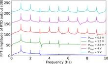
In 1974, physicists first described a device in which electrons pass through an energy barrier thanks to quantum tunneling. Today, that device, the resonant tunneling diode, is becoming the heart of an unusual computing architecture.