Magnetic resonance imaging (MRI) is a non-invasive imaging technique that uses strong magnetic fields and radio waves to create detailed images of the inside of the body, particularly soft tissues like the brain, muscles, and organs. Unlike X-rays or CT scans, MRI does not use ionizing radiation, making it a safer option for repeated imaging. It is a cornerstone of modern medical diagnostics, especially for visualizing brain tumors, spinal cord injuries, joint problems, and cardiac function.
History
The principles of nuclear magnetic resonance (NMR) were discovered in the 1930s and 1940s, but it was not until the 1970s that Paul Lauterbur and Peter Mansfield independently developed the techniques for spatial encoding that made MRI possible. They shared the Nobel Prize in Physiology or Medicine in 2003.
How it works
MRI relies on the magnetic properties of hydrogen nuclei (protons) in water and fat molecules. The patient is placed in a strong magnetic field, which aligns these protons. A radio frequency pulse then knocks them out of alignment. As they relax back to their original state, they emit signals that are detected by coils and processed by a computer to construct a three-dimensional image. Different tissues have different proton densities and relaxation times, which creates contrast in the images.
In practice
MRI is used clinically to diagnose and monitor brain tumors, stroke, multiple sclerosis, spinal cord injuries, and musculoskeletal disorders. In research, it is used to study brain function (fMRI), track the effects of chemotherapy on glioblastoma, and visualize muscle movement in real time.
Fun fact
The magnetic field of an MRI scanner is typically around 1.5 to 3 teslas, which is roughly 30,000 to 60,000 times stronger than Earth's magnetic field.
A courtroom interpreter catches every word, every breath, every whisper in the corner. They have no right to miss a detail, because the meaning of the whole conversation depends on it. Quantum state tomography works in much the same way: it reconstructs the full quantum state of a system from a set of measurements, leaving no detail out.
A sculptor looks at a block of marble and sees the figure within, waiting to be freed from excess material. Instead of adding clay, he removes everything that is not part of the form. The authors of a new study on magnetic resonance took a similar approach: rather than complicating the tissue model by adding ever more details, they stripped away the superfluous to reveal the essence.
A doctor looks at a screen and sees two images of the same part of the body. One shows where blood is rich in oxygen, the other shows how fast it rushes through the finest vessels. Previously, these images were made by different devices, and combining them was difficult. Now engineers have built a device that takes both images simultaneously.
An MRI scanner is a big magnet that aligns hydrogen atoms in the body and then picks up their response signal. Such machines usually take up an entire room and cost as much as a nice house. Engineers from several labs decided to build a scanner that fits in a suitcase and released all the plans as open source.
The coil in an MRI picks up radio signals from hydrogen atoms, like an antenna catching a distant radio station. But the signal is weak, and if the amplifier itself is noisy, the image comes out grainy.
Two scans of the same brain taken at different times are like photographs of the same house before and after renovation. To see what changed, you overlay them and look at the differences. But an MRI scanner is not a perfect camera: it adds its own distortions, as if the photo were taken through slightly warped glass.
A doctor spends years learning to recognize diseases from brain scans. First, they excel at spotting signs of aging, then they master tumors, and suddenly they start confusing what they knew before. Many neural networks behave much the same way: new knowledge overwrites the old.
A magnetic mirror is a tube open at both ends. The magnetic field inside is arranged so that hot plasma doesn't leak out: at the ends, the field lines bunch up like a cork in a bottle. Plasma particles flying toward the exit are repelled by these "corks" and bounce back. But some still escape: that's the classic "loss cone" argument.
Imagine a kinked garden hose: water meets resistance, and pressure drops. A similar process occurs in arteries when they lose their straight geometry. Vascular tortuosity is not just a cosmetic defect but a factor that disrupts hemodynamics and poses a threat of ischemic stroke.
Tomographs don't produce ready-made pictures. They collect numerous 'shadows' — projections of innards from different angles or using various signals — and mathematically reconstruct a 3D picture from them.
Fast radio bursts — millisecond radio pulses arriving from distant galaxies — long remained a puzzle. Their colossal energy, compressed into instants, pointed to cataclysms on neutron stars, perhaps pulsars or magnetars.
Dozens of copper coils in an MRI scanner pick up the faint response from tissues, like microphones trying to catch a whisper in a noisy hall. Each microphone drags a thick bundle of wires behind it. The wires press on each other, create electrical interference, heat up, and add risk for the patient.
The Universe is less than a billion years old, yet at its centers already sit black holes with masses of a billion suns. According to the conventional picture, they grew by voraciously devouring matter, but calculations show that 'feeding' must have been almost continuous, while visible quasar flares last only a few million years. So where does all the accreting fuel go?
The brain doesn't learn every task from scratch: it transfers experience from one activity to another. But how do you know which tasks are truly related? Neuroscientists came up with a clever trick: let an artificial network "peek" at brain activity during one task and try to predict activity during another. If it works well, the tasks are close.
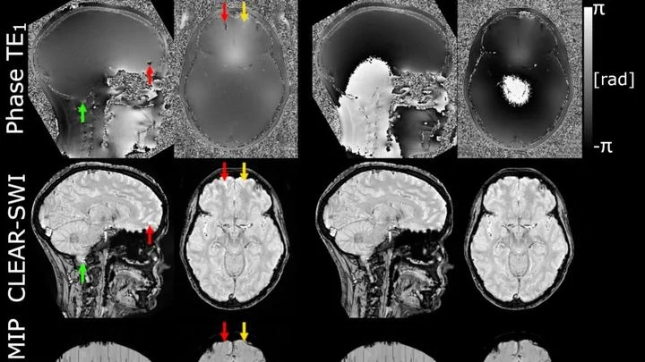
The powerful magnet of an MRI scanner induces weak electric currents in tissues. Their phase behaves like a compass needle trembling with every gust of wind — the slightest disturbance turns the map into a ripple, where instead of small vessels or iron deposits, all you see is noise.
Every second, about a hundred invisible particles, muons, pass through your body. They are born high in the sky when cosmic rays collide with air atoms, raining down on Earth. These particles are so penetrating that they can pass through a multi-story building, but they slightly change direction when they encounter dense material, such as lead or uranium.
As a person breathes, the liver glides smoothly up and down. When treating a tumor with radiation, this motion must be accounted for: the beam must hit the target, not healthy tissue. Usually, breathing is "captured" using special computed tomography (CT), taking a series of frames over one breath cycle.
Photographing a delicate spiderweb trembling in the wind: that's roughly what it takes to look into the blood vessels of a living brain. Ultrasound localization microscopy (ULM) turns ordinary ultrasound into a tool capable of seeing individual capillaries, but only if you get the settings just right.
Drop two pebbles into a pond at the same time — the ripples will spread evenly and in sync. The same happens in a quantum interferometer: a particle is split into two waves that travel along different paths and then meet. Ideally, their crests align.
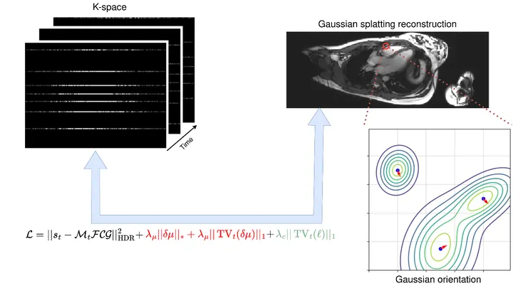
A photographer shoots a runner with a long exposure and gets a blurry silhouette. That's roughly how a standard MRI scanner "sees" moving organs: the heart contracts, the lungs breathe, and the image smears. To avoid this, patients are asked to freeze and hold their breath. But for a child or someone short of breath, that's nearly impossible.
The method called GRASP tackles the problem differently.
The scene is lit by spotlights, but the most interesting action happens in the shadows — where molecules invisible to the eye are hiding. The new camera picks up the heat of these "invisible" objects and turns it into ordinary light.
A proofreader checks a manuscript before it goes to print. They don't know every possible mistake, but they know exactly what flawless text looks like. Spotting a strange symbol or a blurred word, they immediately circle it in red. That's roughly how the new program for checking medical images works.
When you need to see inside the brain, you usually lie in a tight tube of an MRI scanner or get zapped with X-rays. Both methods are costly, bulky, and not always available.
A doctor studies a brain scan: somewhere in the tangle of tissue, a tumor is hiding. One glance picks out small spots and edges, another takes in the overall shape and location. A person combines both, but across hundreds of scans that gets tedious. A new program does the same thing automatically.
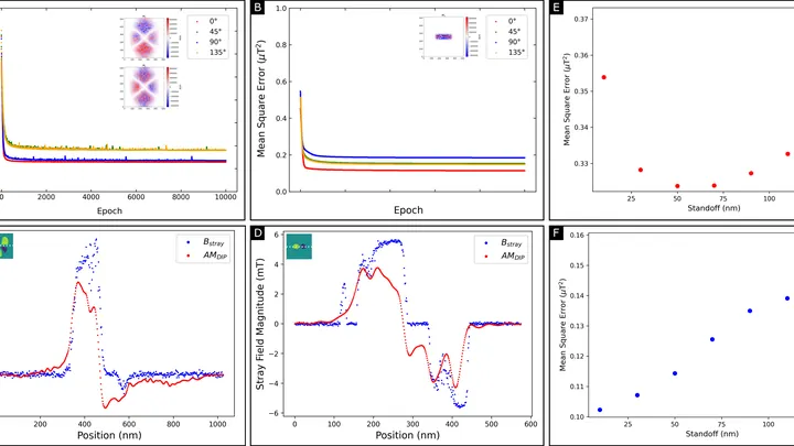
A magnetic film just tens of atoms thick stores information in tiny magnetic vortices. But you can't look inside and see these vortices directly: the instrument only senses the weak magnetic field around the film. Reconstructing the magnetization pattern from such a field is like guessing the shape of an invisible relief from its shadow on a wall.
A doctor rarely makes a diagnosis from a single quick look at a scan. They peer, zoom in, compare with an atlas, flip through articles. Until recently, neural networks worked differently: see the image, immediately produce the text. The authors of the new study decided to give the model time and tools.
A surgeon removes a breast tumor, but how do you know no cancer cells are left at the edge? Right now, the tissue goes to a lab, and the answer comes back days later. If the margin is not clean, a second surgery is needed. The new method promises to give the answer right in the operating room, while the patient is still on the table.
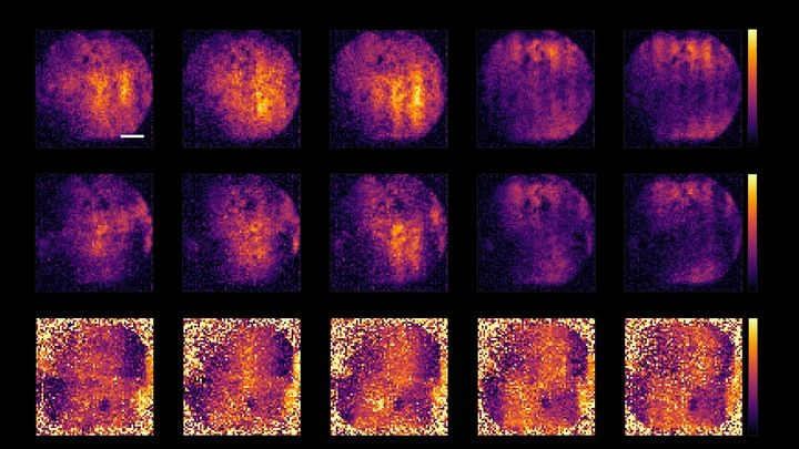
The lungs are not two bags but millions of tiny sacs where air enters and blood flows close by. To see how they work, doctors sometimes use MRI without contrast: the scanner picks up weak signals from the tissues themselves. But these signals are like a radio in a thunderstorm — breathing is irregular, pulse jumps, and the image falls apart.
A regular fridge magnet attracts iron because all its tiny magnetic arrows point in the same direction. But there are materials where the arrows arrange themselves more cunningly: neighboring rows point in opposite directions, so from the outside it looks like there's no magnetism at all. Such materials are called altermagnets.
A weak magnet in an MRI machine is like a dim bulb in a dark room: you get a picture, but details drown in grain. These scanners operate at fields below 0.1 tesla (a typical hospital machine runs at 3 tesla, 30 times stronger), yet they're lightweight, inexpensive, and don't require a special room. The catch: noise eats away at quality.
An MRI scanner is like a camera that photographs your insides, but very slowly. The patient has to lie still for several minutes while the heart keeps beating. If the person moves or the rhythm goes off, the image blurs. Doctors then have to repeat the scan, wasting time and nerves.
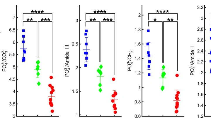
A finger is illuminated with a laser beam, and some of the light returns after traveling inside. That's how Raman spectroscopy works: molecules in bone and skin alter the color of the reflected beam differently, leaving their chemical fingerprints. But skin, muscle, and fat are noisy: their signal drowns out the bone's, like a crowd's chatter overpowering a single singer.
The edge of a magnet works like a guitar fret: a string pressed near the edge sounds different, with a richer set of overtones. Magnetic waves in a thin strip of nickel and iron behave in much the same way.
In a piece of manganese oxide just a few atomic layers thick, electrons behave like a crowd on an escalator: each moves by its own rules, but together they create a common flow. Physicists shone soft X-rays on such a film and knocked electrons out.
In particle scattering experiments, the target is often polarized: nuclei are aligned so that their spins point predominantly in one direction. The degree of this alignment affects the collision outcomes.
A standard MRI machine is a huge magnet that you can't move, plus a powerful computer that turns signals from the body into a picture. In recent years, portable scanners with weaker fields have appeared: they are light, cheap, and can work in a field hospital or an ambulance.
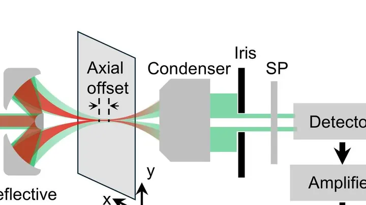
Light we can't see but feel as heat turns out to be the key to cellular life. A new type of microscope uses waves 2000–2500 nanometers long, three times longer than red light, and such waves barely scatter in tissue. Thanks to this, the device peers into a tumor spheroid like a flashlight through frosted glass, illuminating individual molecules.
A fat droplet inside a living cell is like a bobber in rough water. It doesn't move in a straight line but in jerks, sometimes pausing, sometimes speeding up. Previously, such movements could only be seen in a single plane, like watching the bobber from above without knowing its depth.
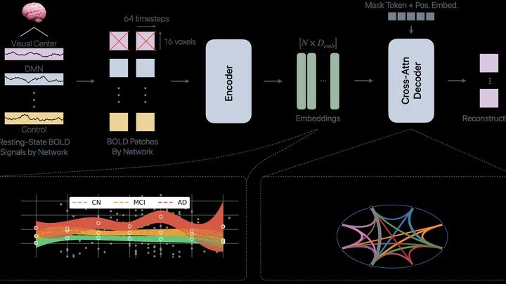
The brain isn't one big computer: it's more like an orchestra, where different groups of musicians play their parts. Neural networks for memory, attention, or emotions are constantly talking to each other. In Alzheimer's disease, this orchestra starts to go off-key: some groups fall silent, others play out of sync.
Every breath in and out shifts internal organs by several centimeters. For an MRI scanner imaging the abdomen, this is like photographing a runner with a long exposure — the image comes out blurred. Clinicians have long known how to deal with this: they ask patients to hold their breath or take a series of fast snapshots between breaths. These methods are simple but effective.