Why the Tumor Doesn't Glow for Everyone express
Imagine a workshop where glowing paint is assembled. A worker takes a precursor, adds the right pigment, and the paint is ready. Nearby, a supervisor redirects some of the paint to another warehouse and dumps the rest as waste. Similarly, brain tumor cells themselves produce the substance that makes them glow under the surgeon's blue lamp. But in some patients, the tumor shines brightly, while in others it barely smolders.
For a long time, the blame was placed on the "supervisors": the pump protein ABCB1 (which exports the glowing substance) and the enzyme FECH (which converts it into another compound). However, when scientists examined tumor samples from over two hundred patients, this hypothesis didn't hold up. No link was found between the activity of the MAPK signaling pathway (the cell's internal command chain) and the pump or the converting enzyme.
Instead, a completely different picture emerged. The more active the MAPK command chain, the less the cell produced of the enzyme PPOX — the very "worker" that converts the precursor into its glowing form. The relationship was inverse: strong signal — weak production, and vice versa. It's as if the workshop manager, instead of worrying about leaks, simply tells the worker to slow down.
If this link is truly causal, then brightness is regulated at the synthesis stage, not at the removal stage. So, to help surgeons see tumor boundaries more clearly, we need to target not the pumps or side reactions, but the assembly process itself. The MAPK command chain becomes the dial we can turn to boost brightness.
Original abstract
as written by the authors arXiv:2608.07865v1
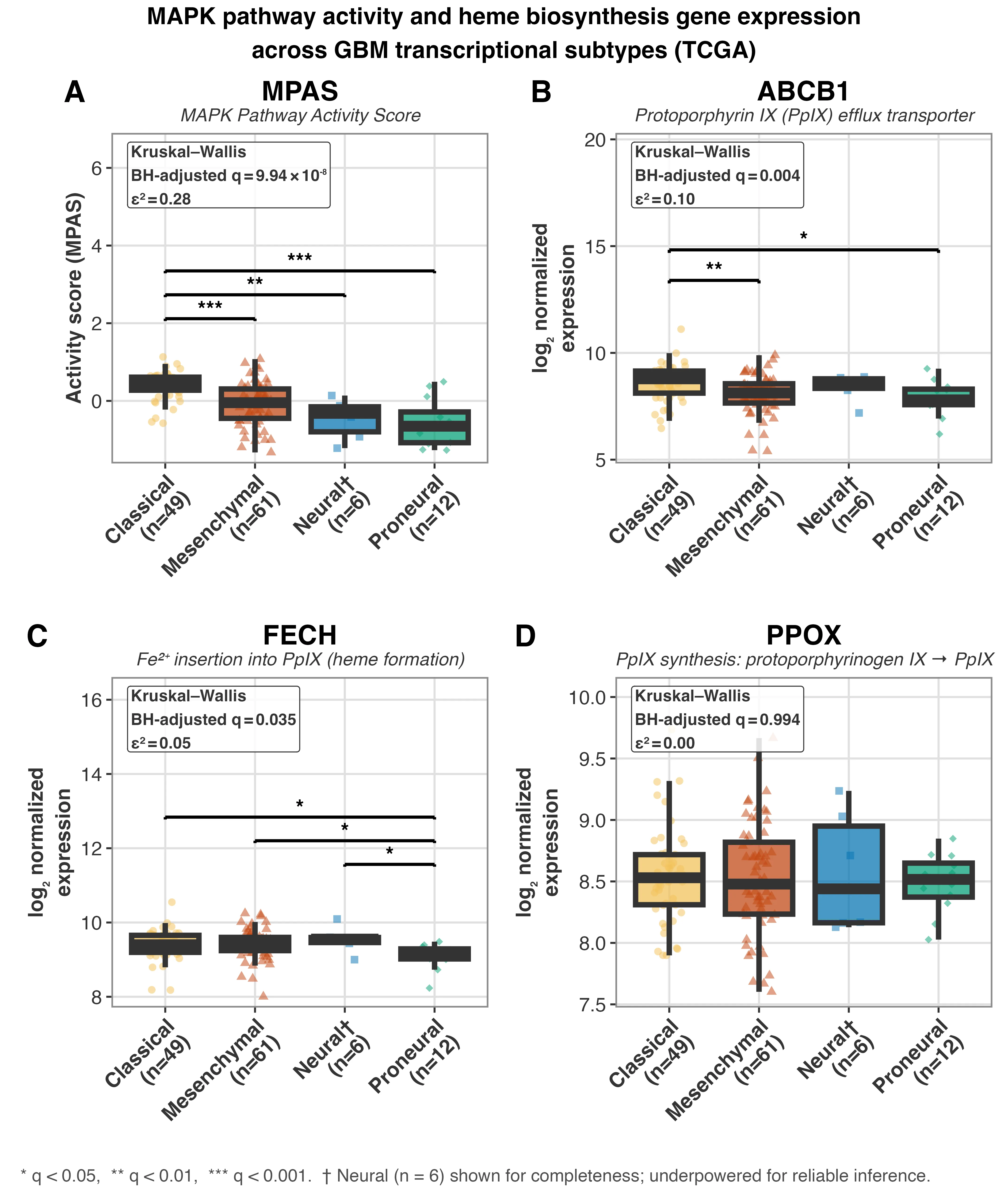
5-aminolevulinic acid (5-ALA) promotes fluorescence-based resection of glioblastoma via protoporphyrin IX (PpIX); however, there is considerable heterogeneity in fluorescence intensity, limiting margin distinction. Cell-line studies show that stimulation of the MAPK pathway results in decreased PpIX levels through increased elimination by ABCB1, an efflux transporter, and ferrochelatase (FECH), the enzyme that converts PpIX to heme. However, the mechanism has yet to be studied in human tissue. Using two independent, purity-adjusted sets of primary IDH-wildtype glioblastoma specimens (TCGA-GBM, n=140, discovery; CGGA, n=87, validation), no reproducible correlation between MAPK signaling and its proposed downstream effectors (ABCB1 and FECH) could be detected. Instead, MAPK activity displayed a reproducibly negative relationship with PPOX, the enzyme that converts protoporphyrinogen IX to protoporphyrin IX: TCGA-GBM (n=133 with purity estimates; rho = -0.40, 95% bootstrap CI [-0.54, -0.25], adjusted p < 0.001), CGGA (rho = -0.28, 95% bootstrap CI [-0.47, -0.07], adjusted p = 0.0342). The finding was robust to permutation testing and to alternative approaches to purity adjustment. Here we report, for the first time, replicated human-tissue evidence for this association. If this correlation reflects causality, MAPK regulation of PpIX would act at the point of synthesis rather than at the clearance steps implied by previous cell-culture studies. We observed differential activation of the MAPK pathway with increased activation in the classical subtype and decreased activation in the proneural subtype. There was no correlation between MAPK pathway activation and overall survival (log-rank p = 0.73). The association between PPOX expression and MAPK pathway activation is biological rather than prognostic, identifying the MAPK pathway as a candidate handle for increasing 5-ALA fluorescence.
The agent 5-ALA that makes tumors glow is essentially a "building block" from which the cell itself assembles the glowing molecule. On its own, it is transparent.