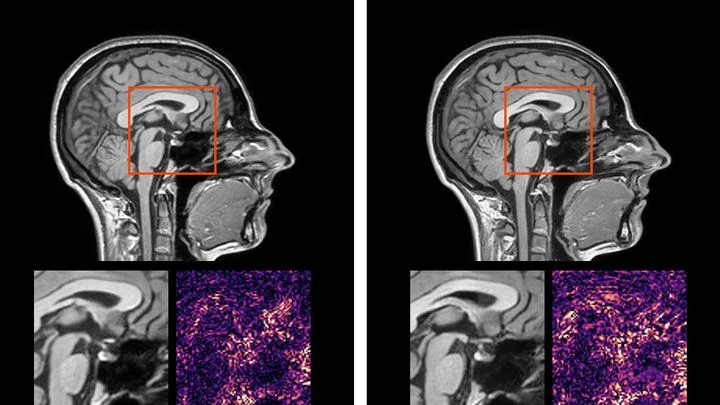
Brain cancer detection using artificial intelligence includes segmentation (delineation) of tumors on MRI images. Convolutional neural networks (CNN) with the U-Net architecture are most effective, processing 3D volumes and classifying each voxel as healthy tissue, necrosis, edema, or tumor. Models are pre-trained on large datasets (e.g., BraTS) with slice-by-slice annotation. The key challenge is distinguishing glioblastoma from metastases and non-tumor formations (abscess, demyelination). Additionally, radiomics (extraction of multiple quantitative features from images) and diffusion-weighted MRI are used.
History
The development of computer-aided brain cancer diagnosis began with expert systems in the 1980s that used thresholding and texture filters. In 1998, the first CAD package for MRI appeared, and in the 2010s, public BraTS (Brain Tumor Segmentation Challenge) competitions accelerated progress. The pivotal moment was the victory of convolutional networks in 2015 (DeepMedic). Since 2018, U-Net-based models and its 3D modifications became the standard. In the 2020s, the FDA approved several AI systems (e.g., icobrain) to assist in clinical decisions, although widespread adoption is hindered by scanner and protocol variability.
How it works
A typical pipeline: (1) preprocessing — intensity normalization, skull stripping, registration to an atlas; (2) an input 3D patch from four channels (T1, T1c, T2, FLAIR) is fed into a U-Net encoder that extracts hierarchical features from edges to texture; (3) the decoder restores spatial resolution with skip connections, outputting probability maps for each class; (4) the loss function is usually a combination of Dice Loss and cross-entropy, accounting for class imbalance; (5) post-processing — removal of small false spots using connected components and morphological operations. To increase robustness, activation consistency is checked under small distortions of the input image (test-time augmentation). The integrated system visualizes the segmentation in the clinician's interface, providing quantitative metrics (volume, radiomics).
In practice
Helps doctors diagnose at an early stage, reduces the number of false alarms, and precisely determines tumor boundaries before surgery or radiation.
Fun fact
Some algorithms can detect a tumor 6–12 months earlier than a radiologist by analyzing barely noticeable changes in signal intensity on consecutive scans.
A stain is found at a crime scene. The expert takes a swab, and a few hours later the instrument outputs a list of proteins that made up the fluid. Previously, a rapid test could be wrong if multiple fluids were present: blood mixed with saliva, and the strip gave a false result.
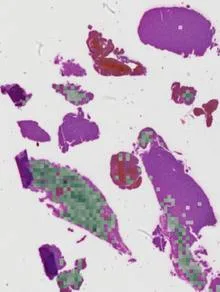
Imagine trying to figure out what happened in a room from the sound that lingers after a gunshot. The shot is gone, but the echo still bounces off the walls. That's roughly how a neuropathologist views a brain tissue section where the tumor itself is absent: the biopsy only captured healthy tissue that reacted to the disease.
A thin section of eucalyptus lies under the microscope objective, and white light, passing through two polarizing filters, turns the wood into a mosaic of purple, yellow, and blue patches. These colors are not random: like letters on a map, they show the direction of the invisible cellulose threads inside the cell walls.
A drop of blood from a fingertip lands on the white membrane of a cartridge. Twenty minutes later, the phone shows whether Lyme disease is present or not. No test tube, no centrifuge, no waiting for days.
Usually, for such a test, blood is first spun in a centrifuge to separate the liquid part — the plasma.
The dummy tips backward in its chair: its head hits the floor. Inside the head, in three Petri dishes, live human nerve cells. They feel the impact just as a real brain would during a fall.
The experiment is simple: the dummy was dropped from a sitting position at angles of 30, 60, and 90 degrees.
High-frequency sound focused on a single point acts like an invisible lens gathering sunlight. Inside a tumor, such a lens can raise the temperature to about 72°C in a second, like hot water from a tap, but inside living tissue. Meanwhile, surrounding healthy cells remain almost untouched.
A single blow to the head is bad enough. But what about a series of blows? That's more complicated, and standard computer models of the brain don't account for it: they treat each impact as if it were the first. But the brain isn't a steel ball: it remembers past strain.
Brain tissue is like a rubber band that has been stretched many times.
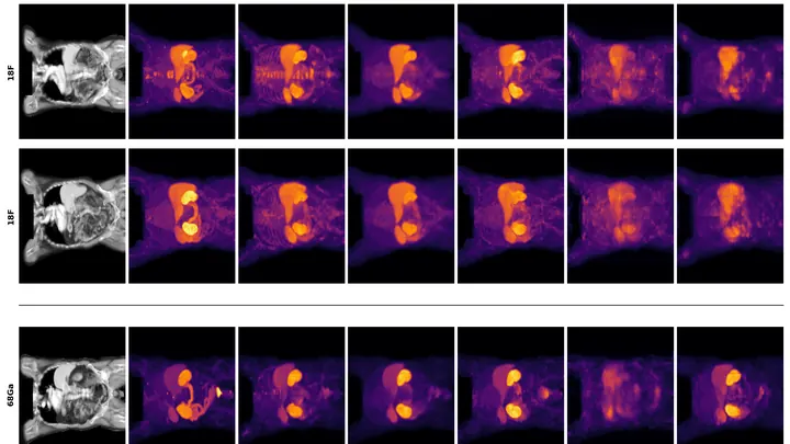
A tumor in a scan is like a tiny bright dot on a vast dark canvas. That dot is what the doctor needs, not a perfectly smooth background. But when a neural network learns to turn a regular CT scan into something resembling a PET scan, it often tries to make everything look nice, and ends up smearing that dot.
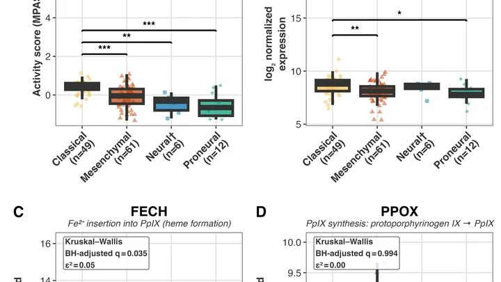
Imagine a workshop where glowing paint is assembled. A worker takes a precursor, adds the right pigment, and the paint is ready. Nearby, a supervisor redirects some of the paint to another warehouse and dumps the rest as waste. Similarly, brain tumor cells themselves produce the substance that makes them glow under the surgeon's blue lamp.
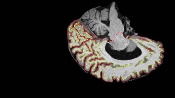
Two scans of the same brain taken at different times are like photographs of the same house before and after renovation. To see what changed, you overlay them and look at the differences. But an MRI scanner is not a perfect camera: it adds its own distortions, as if the photo were taken through slightly warped glass.
A doctor spends years learning to recognize diseases from brain scans. First, they excel at spotting signs of aging, then they master tumors, and suddenly they start confusing what they knew before. Many neural networks behave much the same way: new knowledge overwrites the old.
A flashlight shines through frosted glass: the rays bend, lose brightness, and take different paths. That's roughly how high-intensity ultrasound passes through the skull to the brain. For years, calculating this path required bulky computer models that 'chew' on data for hours.
A bell hidden in a thick box can be made to ring without opening the lid. Just send a sound wave through the walls, and the clapper will tremble. That's roughly how transcranial ultrasound works: sound passes through the skull bones and makes neurons deep in the brain 'ring'—generating electrical impulses.
The proton beam is like a sniper's bullet: it delivers most of its energy at a precisely set point, barely touching tissues on the way in. But over 4–6 weeks of head and neck tumor treatment, the target shifts: the patient loses weight, the tumor shrinks, posture changes. The sniper aimed at one spot, but the target has already moved.
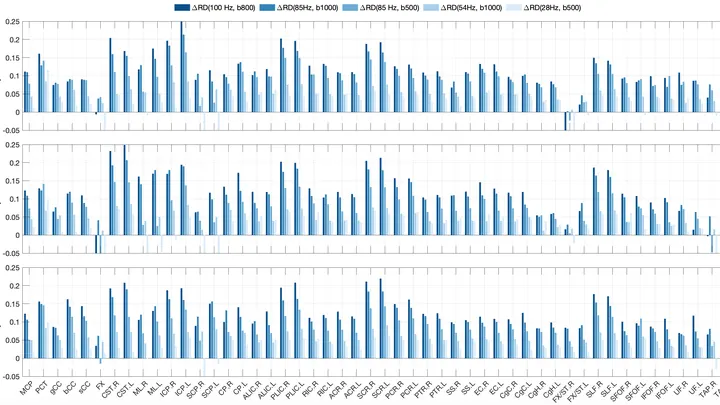
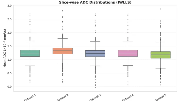
Water inside nerve fibers doesn't move like it does in a glass: it constantly bumps into the walls of axons (the long projections of nerve cells). If you make the pause between MRI radio pulses very short, water doesn't have time to travel far, and its movement reflects the tightness of the surrounding space.
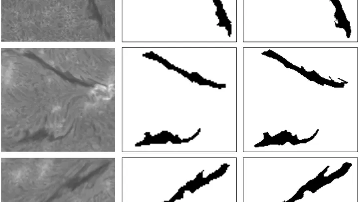
Each morning, dozens of telescopes swing toward the Sun. Their cameras catch a narrow band of red light — the Hα line, discovered by Johann Balmer. The images reveal dark, whimsically curved threads. They look like stitches in a giant tapestry sewn through the blazing corona.
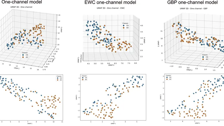
The brain is an orchestra where each group of instruments must come in on time. In Alzheimer's disease, the musicians lose their notes: connections between brain regions get tangled, and the harmonious performance turns into cacophony. Scientists have long seen these disruptions on scans, but they didn't know if the situation could be fixed by touching just one string.
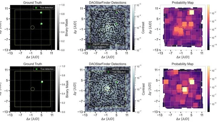
A pebble on a streambed lies still as sunlight sparkles dance on the water's surface. In astronomy, the contrast is similar: a real exoplanet is the stone, while myriad speckles (patches of scattered starlight) are the ripples trembling in instruments.
To capture every detail of a statue's shape, you could break it into millions of tiny cubes and describe each one — slow, heavy, and your computer's memory would groan at the seams. That's how 3D medical scans are usually handled: a CT or MRI is a huge array of such cubes (voxels), and to create or improve an image, the machine has to plow through all of them.
Ordinary AI looks at medical images like puzzles made of unfamiliar pieces: it considers any two unmatched pictures completely different. It's like trying to tell twins apart without knowing they're related. In medicine, however, patients with the same condition look nearly identical on MRI, and the algorithm mistakes 'relatives'.
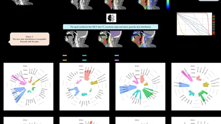
Each morning, a navigator plots a route, bypassing traffic jams and roadblocks. Similarly, before a radiation therapy session, a new AI agent recalculates the irradiation path.
Ultrasound waves bounce off organs to draw their outlines, but on an ovarian scan it's hard to tell a harmless fluid-filled cyst from a solid cancerous mass. Even specialists draw the boundaries differently, and this directly affects treatment choices.
The OvAi Focus program tackles this by learning from thousands of examples.
In boron neutron capture therapy (BNCT), the patient receives an injection of the isotope boron-10, which selectively accumulates in tumor cells. The tumor area is then irradiated with a beam of thermal neutrons.
Medical scans are maps of tissue density. The brightness of each pixel reveals what’s there, much like photometry measures light intensity. But conventional algorithms analyze pixels in isolation, producing jagged organ boundaries.
A new approach compels the program to prioritize smoothness.
Assessing image quality is a job for a meticulous inspector with a toolbox. The IQA-T1 neural network acts exactly like that: it doesn't just stare at the picture, but applies either a spectroscope or a light meter.
To find an epileptic seizure in brain activity recordings, scientists used an approach akin to picking out sharp sounds in a noisy room: instead of deciphering the entire chorus of brainwaves (EEG), they isolated only the strongest spikes.
X-ray magnetic circular dichroism is a precise tool that has long allowed the separation of spin and orbital magnetic moments. However, the classical approach resembles judging an orchestra by its volume: it relies on sum rules, reducing the entire fine structure of the spectrum to a couple of integrals.
Prostate MRI scans often look like photos taken through a dirty window: noise, blurred edges, curvature from field imperfections. Scientists checked what happens if you 'wash' that window: remove the excess, correct the distortions, and accurately align different types of scans.
In the brain's white matter, the signal travels along axons — long neuronal processes wrapped in an insulating myelin sheath. Water fills the space inside axons and outside, in the narrow gaps between fibers.
Dozens of copper coils in an MRI scanner pick up the faint response from tissues, like microphones trying to catch a whisper in a noisy hall. Each microphone drags a thick bundle of wires behind it. The wires press on each other, create electrical interference, heat up, and add risk for the patient.
A radiologist stares at gray-white smudges on the screen — dozens of body slices where every suspicious spot must be found. Manual segmentation of one patient takes half an hour, and a large clinic sees hundreds of such patients. Fatigue and varying experience inevitably introduce variability: two specialists will outline the same lesion slightly differently.
60 mice, 1,980 samples, 234 lung cancer patients. Behind each number lie thousands of genes. Their activity is measured to understand the disease. But the data aren’t uniform: for one patient, you assess a protein level (a continuous number), make a diagnosis (yes/no), and determine the tumor subtype (a category).
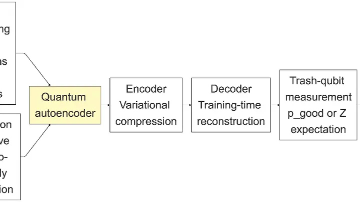
A cook spends years honing the skill of reducing a recipe to three or four essentials and recreating the dish from them. If the ingredients are familiar, the expected meal lands on the plate. A foreign ingredient disrupts compression, and you get something unrecognizable. The Quantum Compressor does the same with medical scans.
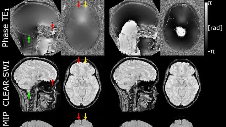
The powerful magnet of an MRI scanner induces weak electric currents in tissues. Their phase behaves like a compass needle trembling with every gust of wind — the slightest disturbance turns the map into a ripple, where instead of small vessels or iron deposits, all you see is noise.
When the canary in the coal mine falls silent, the miners know: the gas is already here, time to get out. Tumor DNA floating in the blood behaves much the same way. The drug bears down on the cancer, and the level of this DNA drops to nearly zero. The test stops seeing it. The doctor sees 'clean' and breathes easy.
But silence can be deceptive.
As a person breathes, the liver glides smoothly up and down. When treating a tumor with radiation, this motion must be accounted for: the beam must hit the target, not healthy tissue. Usually, breathing is "captured" using special computed tomography (CT), taking a series of frames over one breath cycle.
Photographing a delicate spiderweb trembling in the wind: that's roughly what it takes to look into the blood vessels of a living brain. Ultrasound localization microscopy (ULM) turns ordinary ultrasound into a tool capable of seeing individual capillaries, but only if you get the settings just right.
Deep inside the detector at the Large Hadron Collider, protons collide, and for a fleeting moment something heavy is born. It instantly shatters into two Higgs bosons — particles that, like nesting dolls, each hide two pairs of b-quarks.
A smartphone camera captures everything indiscriminately, but the eye immediately picks out what matters. The retina doesn't just pass the image to the brain: it highlights contours on the spot, suppresses noise, and adjusts to brightness.
An engineer building a bridge cannot strengthen one beam without affecting the load on adjacent ones. Drug development works similarly: a molecule must fit snugly into the 'pocket' of a target protein, like a key in a lock, while also being safe, stable, and convenient to administer.