How the Bladder Squeezes Without Paint express
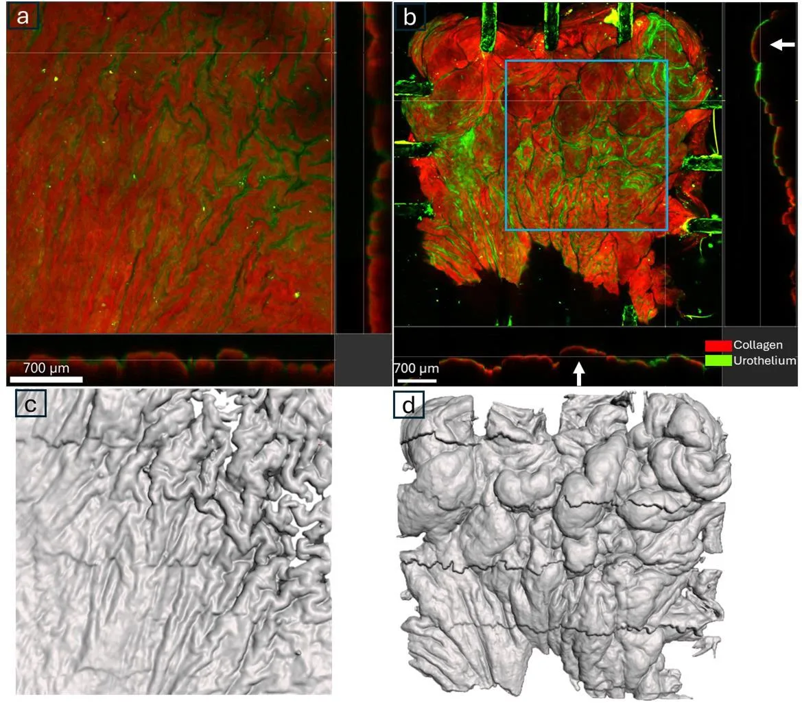
The bladder wall is not a rubber balloon. When the organ empties, its surface wrinkles like crumpled paper, and tension varies from place to place. Previously, to measure this, researchers dotted the tissue with marker spots. But the paint itself changes how living tissue behaves, like an extra layer of tape on a balloon.
The new method skips the paint. A camera captures the natural pattern on the bladder's inner surface, and a program called CoTracker3 follows every tiny patch like a seasoned tracker reading footprints in the sand. The method was tested on four rats: during contraction, the bladder squeezed more along its length than across. Under the microscope, large folds were visible forming.
This contact-free view is useful beyond the bladder. Any material that wrinkles, stretches, and changes shape can be studied more precisely — from heart muscle to plastic wrap.
A rat's bladder is the size of a pea, yet its wall forms folds that resemble a miniature mountain range when it contracts.