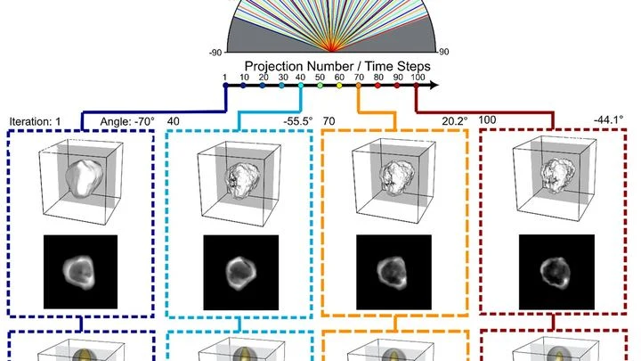
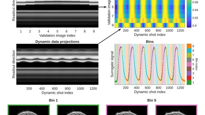
Noninvasive dynamic imaging refers to deep learning methods that capture real-time images of moving organs or tissues without the need for contrast agents, ionizing radiation, or invasive procedures. This is a significant advance because traditional dynamic imaging often relies on contrast dyes (which carry risks) or repeated radiation exposure (which is harmful). By leveraging neural networks, these methods can reconstruct high-quality images from sparse or low-signal data, making it possible to observe physiological motion—like a beating heart or a contracting bladder—safely and continuously.
History
The approach emerged in the mid-2010s with the advent of deep learning in medical imaging, particularly for MRI reconstruction. The specific application to dynamic, contrast-free imaging has been developing rapidly since around 2018.
How it works
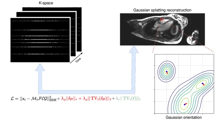
These methods typically use deep learning to either enhance low-quality images or to reconstruct full images from under-sampled data. For example, in MRI, a network can learn to fill in missing k-space data, allowing faster acquisition that captures motion without blurring. In ultrasound or optical imaging, neural networks can separate signal from noise, enabling imaging without contrast agents by detecting subtle intrinsic contrasts like blood flow or tissue density changes.
In practice
It is used in cardiac MRI to image heart motion in real time, and in bladder imaging to study filling and contraction without catheterization or contrast dye.
Fun fact
Some of these techniques can image the bladder's contraction cycle using only natural contrast from urine, eliminating the need for any external agent.
A superconducting detector usually works like an honest ruler: absorb a photon, get a pulse whose amplitude is strictly proportional to the energy. This mode is convenient, but it has a ceiling: a weak signal easily gets lost in the amplifier noise.
Now imagine a swing.
A protein is like a tiny machine that switches on and off on command. The command arrives when another molecule sticks to it, like a key in a lock. The protein then changes shape and passes the signal along. But exactly how the signal travels inside the protein has long been a mystery.
Two scans of the same brain taken at different times are like photographs of the same house before and after renovation. To see what changed, you overlay them and look at the differences. But an MRI scanner is not a perfect camera: it adds its own distortions, as if the photo were taken through slightly warped glass.
Conventional neural networks learn from thousands of examples, burning hours and gigawatts. Physicists have found a quantum workaround. As early as Max Planck showed the discreteness of energy, today this allows generation without training.
A proofreader checks a manuscript before it goes to print. They don't know every possible mistake, but they know exactly what flawless text looks like. Spotting a strange symbol or a blurred word, they immediately circle it in red. That's roughly how the new program for checking medical images works.
Your arm lifts a cup from the shelf — a familiar motion you don't notice until muscles tire. Then the trajectory changes: the elbow shifts slightly, the hand trembles. A doctor usually catches these changes by hand, feeling the patient's muscles.
A voice recorder captures a city square. You hear the hum of a crowd, footsteps, snatches of conversation, the rumble of traffic. Somewhere in that sonic stew, a street musician plays a simple tune on a flute. Hearing it isn't easy: there's just too much mixed together.
A flashlight spreads light in a broad beam, while a laser cuts a narrow path and picks out the finest details. In magnetic resonance imaging (MRI), the gradient, a rapid change in the magnetic field along the body, plays the role of that laser.
The vessels inside the skull resemble a mountain road's hairpin turns — sharp bends, narrowings, unexpected curves. When a traffic jam forms on such a serpentine, blood stops reaching the brain properly. To predict trouble, doctors need to see the entire flow: where it accelerates, where it swirls into eddies. The full picture comes from a special MRI, like slow-motion cinematography of fluid.
You taste a fruit smoothie through two different straws. One lets through only the big chunks, the other only the liquid. Individually, the sensations are misleading, but together they give you the key: here, it seems, is banana, and here, strawberry.
The lungs are not two bags but millions of tiny sacs where air enters and blood flows close by. To see how they work, doctors sometimes use MRI without contrast: the scanner picks up weak signals from the tissues themselves. But these signals are like a radio in a thunderstorm — breathing is irregular, pulse jumps, and the image falls apart.
A tiny gold star melts under the beam of an electron microscope, its points slowly rounding off. In the past, such a process could only be seen in snapshots taken with long pauses, and the probing beam itself would alter the sample.
Plasma is a gas in which electrons are stripped from atoms, behaving like a soup of charged particles. To predict how such a soup flows and heats up, you need to know how particles collide and exchange energy.
The heart is not a perfect sphere; its shape changes with every beat. To build an accurate 3D copy for a computer model, doctors typically take dozens of MRI slices. But that's like assembling a watermelon from thin slices: the fewer slices, the more guesswork.
A new approach teaches a neural network to understand the heart's shape from sparse contours.
An MRI scanner is like a camera that photographs your insides, but very slowly. The patient has to lie still for several minutes while the heart keeps beating. If the person moves or the rhythm goes off, the image blurs. Doctors then have to repeat the scan, wasting time and nerves.
The boundary between two liquids is like an invisible skin: it's there, but you can't touch it with your finger. Scientists have found a way to measure its elasticity without touching it. They made a tiny probe oscillate above the surface, and between the probe and the interface lay a thin film of liquid.
The heart doesn't contract all at once but as a wave: an electrical signal travels through the muscle, and behind it, like ripples spreading on water, a contraction rolls through. In arrhythmia, this wave goes awry: it stalls in one spot, loops in another, and the pump starts working in a disorganized way.
Light passing through ripples on a water surface paints dancing patterns on the bottom of a pool. Engineers have used a similar idea to make light compute. They fabricated a tiny plate with a wavy relief: each ridge and groove deflects rays in its own way. When an image falls on such a surface, the light transmitted through it converges at different points behind it.
An antibody is a protein that catches viruses and bacteria. When an enemy enters the body, immune cells start trying out different antibody variants, as if testing different keys against a lock. Beneficial changes are kept, harmful ones are discarded. Over a few weeks, this process yields an antibody that fits its target perfectly.
A photographer snaps a runner: to avoid a blurry shot, a short shutter speed is needed. An MRI scanner is like that photographer, but its 'shutter speed' is long — by the time it captures, the muscle has already changed. That's why dynamic MRI (a series of volumetric frames in motion) is a tough challenge: data are scarce, and motion doesn't wait.
A drop of blood lands on a tiny sensor, and within a minute the doctor knows how many different defenders are in it. That's the future of blood testing, brought closer by Danish engineers.
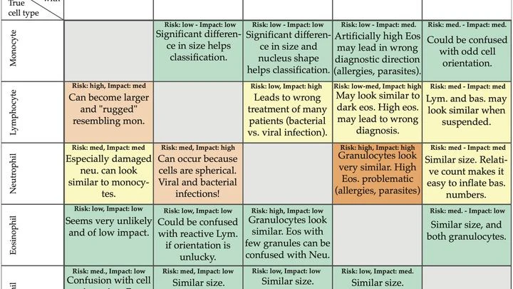
Instead of a regular microscope with lenses, they built a lensless device. A cell flows over the sensor, which records a pattern of dark and light rings — a hologram.
An aneurysm is a weak spot in a blood vessel wall that bulges out like a bubble on an old bicycle inner tube. If the bubble bursts, blood floods into the brain. Doctors need to know how stressed the aneurysm wall is to decide whether to operate or just monitor.
In a dark room, on a table, sit a laser pointer, a piece of glass with tissue, and an ordinary phone camera. Light passes through the sample, and the image shows not cells but a pattern of bright and dark stripes, like the shadow of leaves on a wall.
A pathologist looks through the eyepiece and sees a pink-purple tissue section — the classic hematoxylin and eosin stain. The dyes bind to nuclei and cytoplasm, and the color tells the doctor about the disease. But preparing the slide itself takes hours: fixation, paraffin embedding, sectioning, staining.
This new device skips the dye.
A neural network originally trained to distinguish real gravitational waves from noise in a gravitational wave detector recognizes bearing wear from vibrations without retraining.
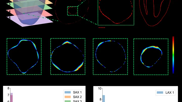
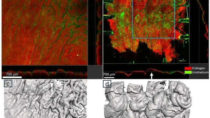
The bladder wall is not a rubber balloon. When the organ empties, its surface wrinkles like crumpled paper, and tension varies from place to place. Previously, to measure this, researchers dotted the tissue with marker spots. But the paint itself changes how living tissue behaves, like an extra layer of tape on a balloon.
The new method skips the paint.
A translator in a conference room hears not only the words but also the echo of previous phrases still bouncing around the ceiling. If they get distracted by every reverberation, an hour of conversation turns into two. Similarly, a complex system, whether an ocean or a power grid, receives random jolts that do not vanish instantly but drag behind them a tail of memory.
A translator learns not from a dictionary but from speech. They listen to native speakers and pick up rules that are written in no textbook. Similarly, a new method for forecasting complex systems such as climate or turbulence does not require knowing their equations. Instead, it looks at data and learns to predict their behavior.