A Map of Brain Aging express
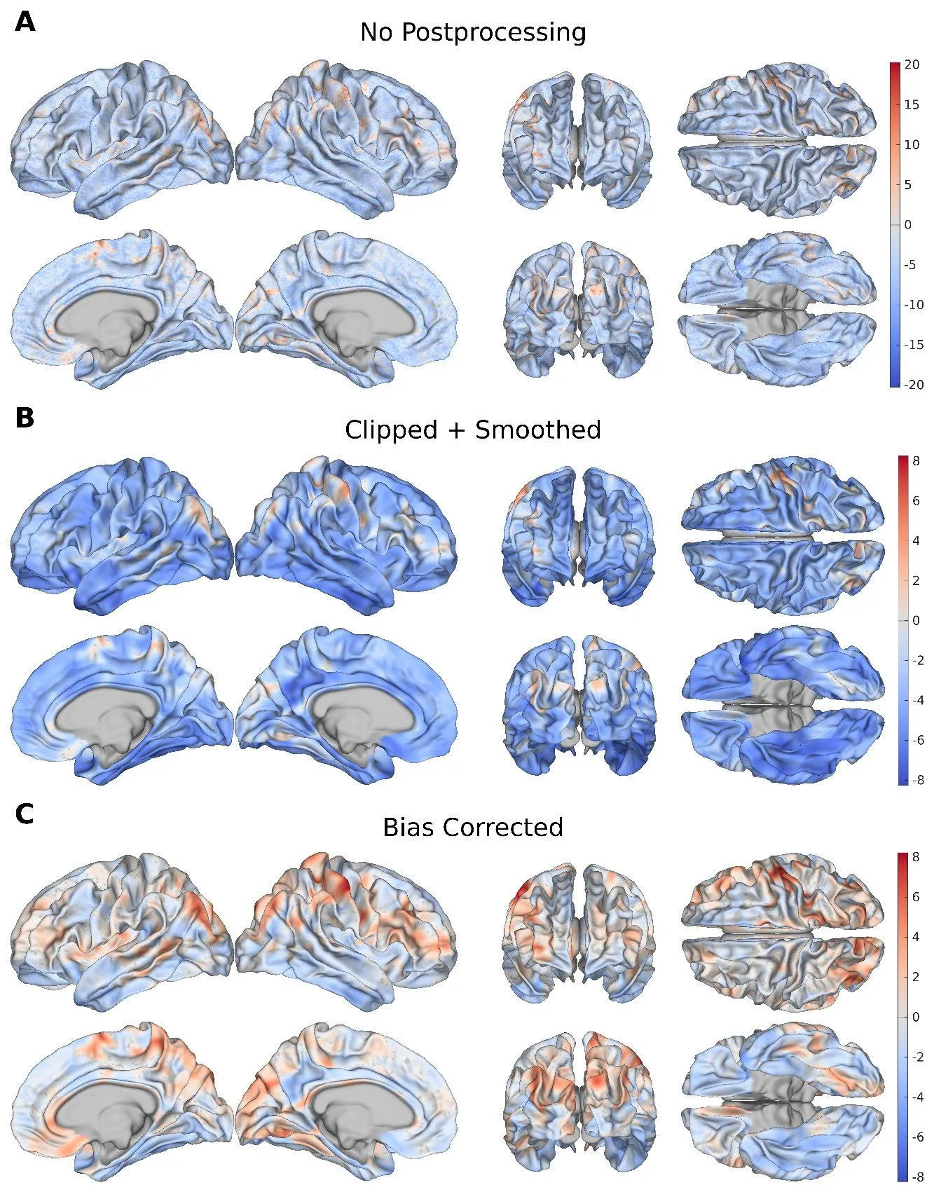
The cerebral cortex is like a crumpled sheet of paper: it has thickness, curves, and folds of varying depth. With age, this sheet changes unevenly — thinning in some places, losing the sharpness of its folds in others. Previously, doctors assessed brain age as a whole, as if looking at the sheet from a distance. Now, a neural network examines every point on the surface with a step of 1.37 mm, like a magnifying glass that reveals the aging of individual regions.
The model was trained on scans from 14,423 healthy adults. It found that in healthy people, the fastest-aging areas are those that connect different brain regions — the association areas of the cortex. With mild memory impairment, aging spreads more widely, involving the parahippocampal gyrus, a region important for memory formation. In Alzheimer's disease, age-related changes affect almost the entire cortex, especially the temporal lobes.
The difference between the actual brain age and the predicted one correlates with tests of memory and thinking. The more a cortical region 'looks older,' the worse a person performs on tasks. This means that a map of local aging is not just a picture but a tool for early diagnosis.
The neural network views the cortex as a network of roads: each point is connected to its neighbors, and the aging of one area pulls others along. This approach allows us to see where breakdown begins long before obvious symptoms appear.
Original abstract
as written by the authors arXiv:2601.10912
Estimating brain age (BA) from T1-weighted magnetic resonance images (MRIs) provides a powerful framework for quantifying anatomical brain aging. Whereas global BA (GBA) summarizes overall brain health, local BA (LBA) provides cortically specific patterns of aging at the subject level. Although previous studies have examined anatomical contributors to GBA, to our knowledge, no framework has been established to estimate LBA using cortical morphology. To address this gap, we introduce a graph neural network (GNN) that uses morphometric features$$\unicode{x2013}$$cortical thickness, surface area, curvature, gray/white matter intensity ratio (GWR), sulcal depth$$\unicode{x2013}$$to estimate LBA across the cortical surface at high spatial resolution (mean inter-vertex distance = 1.37 mm). Trained on cortical surface meshes extracted from the MRIs of cognitively normal (CN) adults (N = 14,423), our model achieves lower mean absolute error (MAE) than the existing state-of-the-art while identifying more biologically plausible patterns of aging in Alzheimer's disease (AD) on the ADNI dataset. Association cortices emerge as primary sites of morphometric aging in CNs, whereas mild cognitive impairment is characterized by widespread aging that is pronounced in the parahippocampal gyrus. AD subjects demonstrate significant aging across the entire cortex, particularly within medial temporal regions and associated cortical networks. Feature ablation highlights curvature and GWR as preferentially sensitive to AD pathology. Regional LBA gaps are significantly associated with neuropsychological measures of AD-related cognitive impairment, linking cortical aging patterns to clinical outcomes. These results demonstrate that GNN-based modeling of cortical morphometry enables biologically interpretable mapping of local brain aging with greater interpretability than prior work.
The average distance between points on the cortex map is 1.37 mm — thinner than a matchstick.
In the series 'Altered Carbon,' brain age could be read directly: this work brings such diagnostics closer.