A brain scan is any technique that produces images of the brain's structure or function. Structural methods like MRI and CT reveal anatomy, while functional methods like fMRI, PET, and EEG show brain activity. Brain scans have revolutionized neuroscience, allowing researchers to link cognitive processes to specific regions and clinicians to diagnosetumors, strokes, and neurodegenerative diseases.
History
The first human brain CT scan was performed in 1971 by Godfrey Hounsfield, and MRI followed in the late 1970s. fMRI was developed in 1990 by Seiji Ogawa, who discovered the BOLD contrast mechanism.
How it works
MRI uses strong magnetic fields to align hydrogen nuclei in water molecules, then radiofrequency pulses perturb them; as they relax back, they emit signals that encode tissue properties. fMRI detects blood-oxygen-level-dependent (BOLD) signals, since active brain regions consume more oxygen and attract more blood flow. PET scans inject a radioactive tracer that accumulates in metabolically active areas, emitting positrons that annihilate with electrons, producing gamma rays detected by a ring of sensors. EEG measures electrical activity via electrodes on the scalp, capturing millisecond-scale dynamics but with poorer spatial resolution.
In practice
Brain scans are used clinically to diagnose stroke, tumors, and multiple sclerosis, and in research to map neural connectivity, study memory, and develop brain-computer interfaces.
Fun fact
A single fMRI scan can generate over 100,000 data points per second, requiring powerful supercomputers to reconstruct the final image.
Old wiring in a house doesn't announce itself right away: the bulbs still glow, but the light has dimmed, and switches sometimes respond with a delay. Neuromuscular junctions age in much the same way. These are the contacts through which a nerve tells a muscle fiber to contract.
Imagine trying to figure out what happened in a room from the sound that lingers after a gunshot. The shot is gone, but the echo still bounces off the walls. That's roughly how a neuropathologist views a brain tissue section where the tumor itself is absent: the biopsy only captured healthy tissue that reacted to the disease.
In a print shop, every page of a book used to be set by hand. One mistake, and the whole print run is at risk. RNA sequencing of the brain looked much the same until recently: each sample demanded hours of manual work, and short reads, like torn scraps of pages, could not be pieced together into the full text of a gene. Now there is a production line that reads every page from start to finish.
A radiologist looks at an abdominal scan and sees not just organs but their spatial relationships: which kidney is larger, where the tumor lies relative to a vessel. It's like reading a map, where it's not enough to know where a city usually stands; you also need to measure the distance between two points on the ground.
In 2023, in Belgium, 101 people aged 50 to 69 spent several days in a lab where their brains were scanned, their sleep was recorded, and they were asked to solve memory and attention tasks. That is how the COFITAGE dataset was born, describing each participant from five angles: molecular, structural, physiological, genetic and cognitive.
In a school report card, each grade speaks for itself, but you can only tell how a student is doing overall by looking at the full report. For drugs that act on the brain, such a report card was missing for a long time. Every test on a miniature brain grown in a dish gave its own signal: some genes switched on, others switched off, electrical impulses sometimes sped up and sometimes went quiet.
Every heartbeat sends a wave through the cerebral arteries that not only pushes blood but also briefly inflates the vessel. The arterial wall, like a tightly stretched rubber tube, passes that motion to the neighboring brain tissue, and the brain pulses ever so slightly in time with the heartbeat.
A doctor looks at a screen and sees two images of the same part of the body. One shows where blood is rich in oxygen, the other shows how fast it rushes through the finest vessels. Previously, these images were made by different devices, and combining them was difficult. Now engineers have built a device that takes both images simultaneously.
Two scans of the same brain taken at different times are like photographs of the same house before and after renovation. To see what changed, you overlay them and look at the differences. But an MRI scanner is not a perfect camera: it adds its own distortions, as if the photo were taken through slightly warped glass.
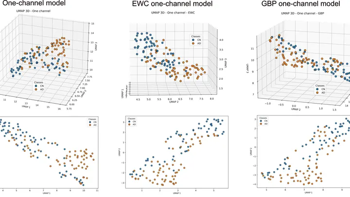
A doctor spends years learning to recognize diseases from brain scans. First, they excel at spotting signs of aging, then they master tumors, and suddenly they start confusing what they knew before. Many neural networks behave much the same way: new knowledge overwrites the old.
A bell hidden in a thick box can be made to ring without opening the lid. Just send a sound wave through the walls, and the clapper will tremble. That's roughly how transcranial ultrasound works: sound passes through the skull bones and makes neurons deep in the brain 'ring'—generating electrical impulses.
One hundred milliseconds. In that time, after a lightning strike or a short circuit, the power system either returns to normal operation or loses stability. A new-generation Neural Network Oscillations learns to sense this moment, like an experienced tightrope walker anticipating a gust of wind.
Medical scans are maps of tissue density. The brightness of each pixel reveals what’s there, much like photometry measures light intensity. But conventional algorithms analyze pixels in isolation, producing jagged organ boundaries.
A new approach compels the program to prioritize smoothness.
Tomographs don't produce ready-made pictures. They collect numerous 'shadows' — projections of innards from different angles or using various signals — and mathematically reconstruct a 3D picture from them.
Dozens of copper coils in an MRI scanner pick up the faint response from tissues, like microphones trying to catch a whisper in a noisy hall. Each microphone drags a thick bundle of wires behind it. The wires press on each other, create electrical interference, heat up, and add risk for the patient.
Studying distant exoplanets, scientists look for signs of life — for instance, water and carbon — and signs of technology: radio signals, megastructures. But between these clues there is a yawning gap. If intelligence hasn't created machines or they've vanished, we won't notice it.
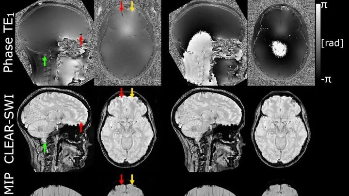
The powerful magnet of an MRI scanner induces weak electric currents in tissues. Their phase behaves like a compass needle trembling with every gust of wind — the slightest disturbance turns the map into a ripple, where instead of small vessels or iron deposits, all you see is noise.
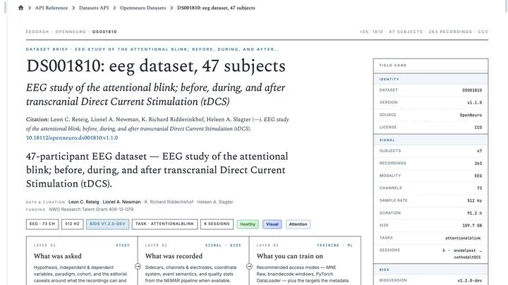
The open archives OpenNeuro and NEMAR hold 791 recordings of the brain's electrical activity from nearly forty thousand people. In total, that's 86,051 hours of signals — roughly ten years of continuous listening. Previously, to take even one such recording off the shelf, you had to write thousands of lines of code: download, unzip, fix format errors, and slice it into chunks of the desired length.
When the conductor leaves the orchestra, the musicians keep playing from memory for a while. But without a unified rhythm, each one starts to speed up or slow down on their own, and the harmonious symphony turns into disjointed noise. According to new research, the brain and heart behave much the same way in the last hours of a person's life.
Photographing a delicate spiderweb trembling in the wind: that's roughly what it takes to look into the blood vessels of a living brain. Ultrasound localization microscopy (ULM) turns ordinary ultrasound into a tool capable of seeing individual capillaries, but only if you get the settings just right.
A smartphone camera captures everything indiscriminately, but the eye immediately picks out what matters. The retina doesn't just pass the image to the brain: it highlights contours on the spot, suppresses noise, and adjusts to brightness.
A photographer shoots a runner with a long exposure and gets a blurry silhouette. That's roughly how a standard MRI scanner "sees" moving organs: the heart contracts, the lungs breathe, and the image smears. To avoid this, patients are asked to freeze and hold their breath. But for a child or someone short of breath, that's nearly impossible.
The method called GRASP tackles the problem differently.
For one person, the disease begins with a tremor in the hand — the cup dances on its saucer as they carry it to the table. For another, the legs suddenly refuse to move smoothly: the step becomes short and shuffling, as if the floor were suddenly covered in a sticky layer. These are not two different diseases but two faces of the same one — Parkinson's disease.
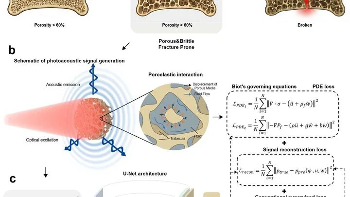
A laser beam strikes bone, and it responds with a nearly inaudible ultrasonic echo. Hidden in that sound is information about every microcrack and every fluid-filled pore. The trouble is that conventional computer programs don't understand this language: they see the signal but don't know the physics that produced it.
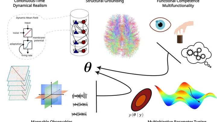
Engineers are building a car. Some assemble an exact replica of the engine, every piston and spark plug, but the car doesn't move: there's no steering wheel or wheels. Others build a fast self-driving go-kart, but its engine is made of playdough and doesn't resemble a real one. This is roughly the situation in brain science.
The hand traces the word "mom" in the air — familiar arcs and strokes. The brain, meanwhile, is not flipping through a photo of each letter. It launches a chain of simple movements.
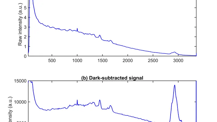
A drop of blood from a vein falls under three beams, and each one drags its own clues into the light. The infrared beam makes molecules vibrate — from that trembling, you learn how much sugar and fat is in the sample. A green laser scatters off proteins and lipids, shifting color — revealing the remodeling of cell membranes.
An X-ray tomograph is like a carpenter who examines a log from all sides to see where the void is inside. It takes dozens of flat shadow images, rotating the source and detector around the object. Then a 'reconstruction' program glues these shadows into a 3D model. The quickest reconstructor is the FBP algorithm (filtered back projection).
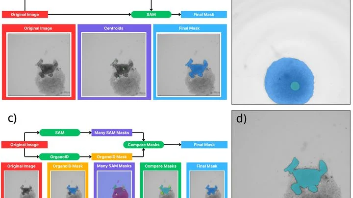
A biologist looks through a microscope at dozens of tiny cell spheres — spheroids. From these, miniature organ-like structures will later grow. The task is similar to a baker's job of checking whether the dough has risen evenly in each mold. Only here, instead of molds, there's a nutrient medium, and instead of dough, there are living cells.
Imagine a river that doesn't flow in an open channel but through the inside of a sponge, with its currents shifting from place to place. That's a good way to think about the brain's glymphatic system: a network of ultra-fine channels around blood vessels, through which cerebrospinal fluid seeps into the tissue and washes away the waste that has piled up during the day.
A doctor studies a brain scan: somewhere in the tangle of tissue, a tumor is hiding. One glance picks out small spots and edges, another takes in the overall shape and location. A person combines both, but across hundreds of scans that gets tedious. A new program does the same thing automatically.
Ultrasound penetrates bone like sunlight through frosted glass, but the image comes out blurry. The skull is uneven and dense, scattering sound waves, so hitting a precise spot in the brain is tricky. Conventional devices try to fix this with electronics, but the result is still far from perfect.
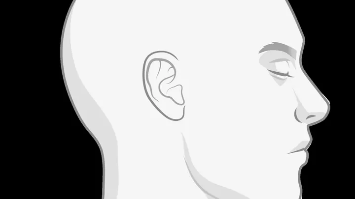
A person hears a ringing that isn't there. The doctor asks them to describe it — loudness, pitch, persistence. But it's subjective: one might say "like a mosquito," another "like steam from a kettle." We need a way to see tinnitus directly, in the brain itself.
Your finger is a living sensor: beneath the skin, arteries pulse in time with your heart. Traditional blood pressure cuffs squeeze your arm and give a single number, but this ring uses eight electrodes to pass a weak current through the tissue and map blood flow.
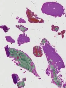
A pathologist stains a tissue section with hematoxylin and eosin — cells turn blue and pink, and under the microscope you can see their shape and arrangement. But which genes are active in each cell cannot be read from such an image. That requires a separate, expensive assay that reveals the activity of thousands of genes at once, but it is rarely performed.
A mole on your skin looks like a mosaic of colored pieces. Each piece is a small patch of the image where color and texture barely change. Such pieces are called superpixels. If you connect them with lines, you get a network that looks like a subway map: stations are the pieces, and lines are the connections between them. The weight of each line shows how strongly two pieces are related.
At night in a dark room, the human body emits a barely perceptible glow. This light is so faint that only highly sensitive detectors can catch it. Some researchers thought this glow from the head could tell us about brain activity — no scanners or wires needed.
But when put to the test, the story changed.
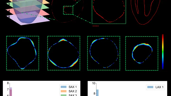
The heart is not a perfect sphere; its shape changes with every beat. To build an accurate 3D copy for a computer model, doctors typically take dozens of MRI slices. But that's like assembling a watermelon from thin slices: the fewer slices, the more guesswork.
A new approach teaches a neural network to understand the heart's shape from sparse contours.
A stroke hits suddenly, but before the strike, the brain sends quiet signals. Blood vessels stop smoothly regulating blood flow, like a thermostat that starts overheating or overcooling a room without reason. This imbalance is invisible on ordinary wearables, but it can be caught by listening to several body rhythms at once.